| Article ID | Journal | Published Year | Pages | File Type |
|---|---|---|---|---|
| 2947038 | Journal of the American College of Cardiology | 2013 | 11 Pages |
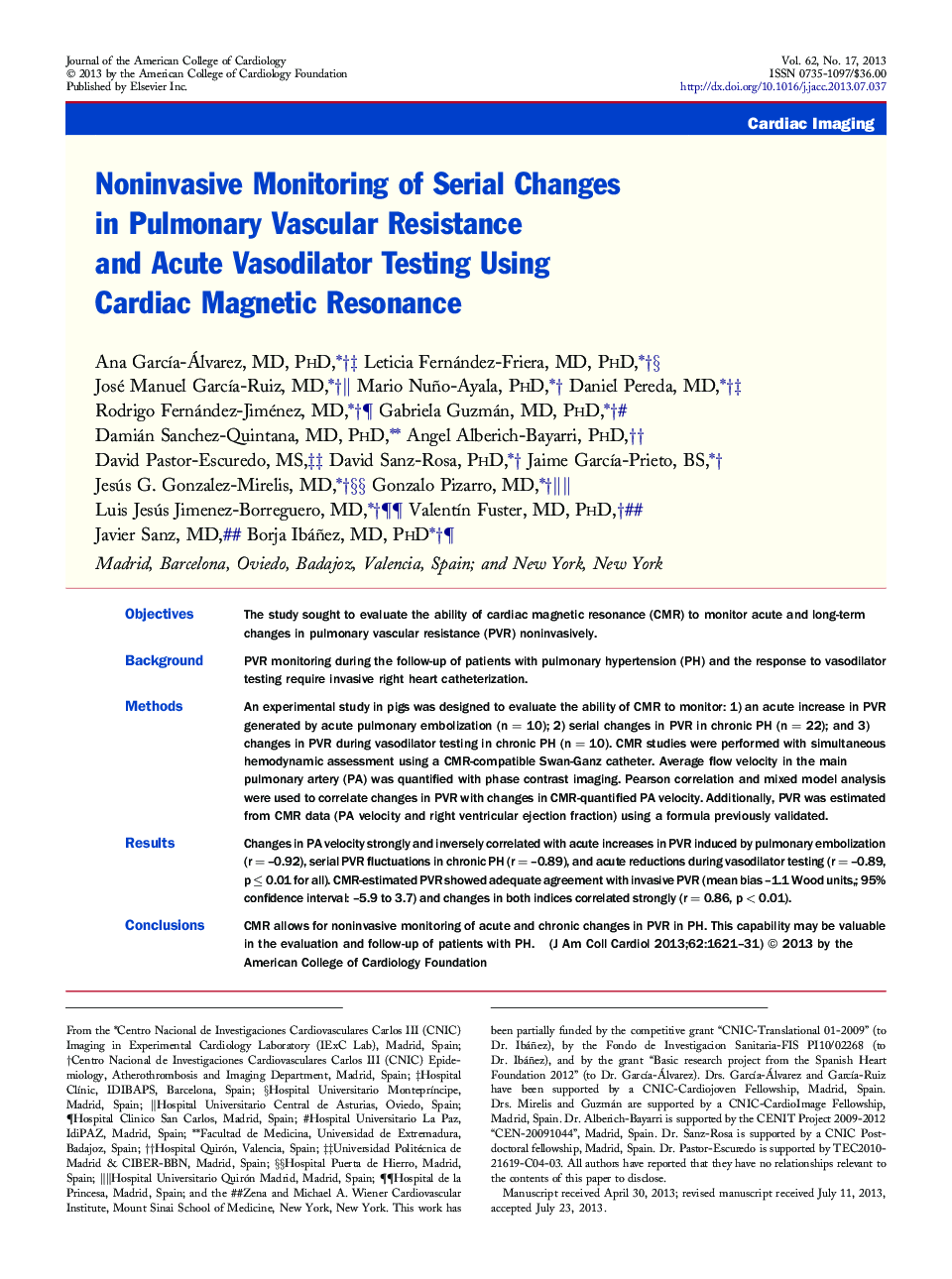
ObjectivesThe study sought to evaluate the ability of cardiac magnetic resonance (CMR) to monitor acute and long-term changes in pulmonary vascular resistance (PVR) noninvasively.BackgroundPVR monitoring during the follow-up of patients with pulmonary hypertension (PH) and the response to vasodilator testing require invasive right heart catheterization.MethodsAn experimental study in pigs was designed to evaluate the ability of CMR to monitor: 1) an acute increase in PVR generated by acute pulmonary embolization (n = 10); 2) serial changes in PVR in chronic PH (n = 22); and 3) changes in PVR during vasodilator testing in chronic PH (n = 10). CMR studies were performed with simultaneous hemodynamic assessment using a CMR-compatible Swan-Ganz catheter. Average flow velocity in the main pulmonary artery (PA) was quantified with phase contrast imaging. Pearson correlation and mixed model analysis were used to correlate changes in PVR with changes in CMR-quantified PA velocity. Additionally, PVR was estimated from CMR data (PA velocity and right ventricular ejection fraction) using a formula previously validated.ResultsChanges in PA velocity strongly and inversely correlated with acute increases in PVR induced by pulmonary embolization (r = –0.92), serial PVR fluctuations in chronic PH (r = –0.89), and acute reductions during vasodilator testing (r = –0.89, p ≤ 0.01 for all). CMR-estimated PVR showed adequate agreement with invasive PVR (mean bias –1.1 Wood units,; 95% confidence interval: –5.9 to 3.7) and changes in both indices correlated strongly (r = 0.86, p < 0.01).ConclusionsCMR allows for noninvasive monitoring of acute and chronic changes in PVR in PH. This capability may be valuable in the evaluation and follow-up of patients with PH.
