| Article ID | Journal | Published Year | Pages | File Type |
|---|---|---|---|---|
| 2951829 | Journal of the American College of Cardiology | 2006 | 7 Pages |
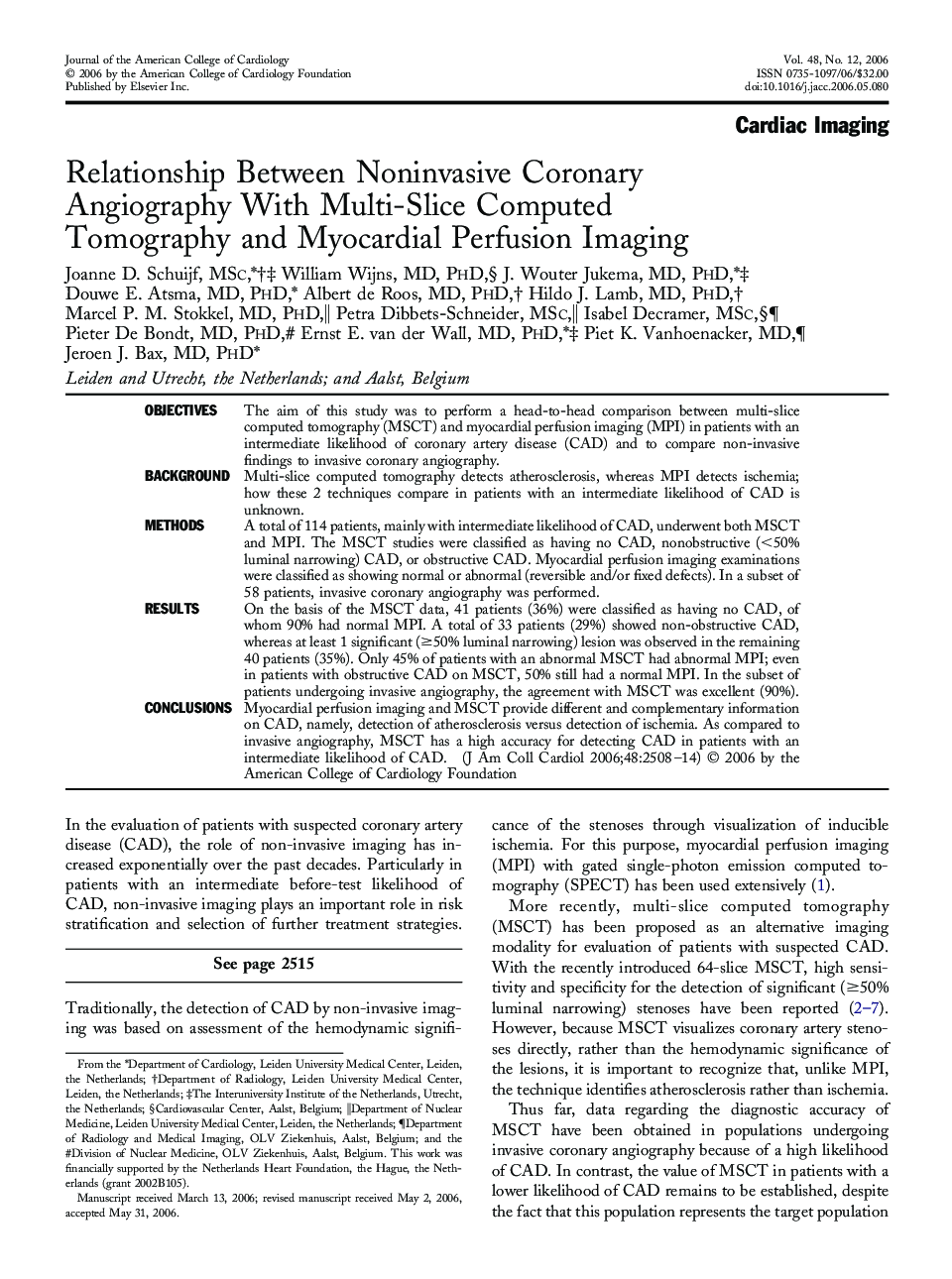
ObjectivesThe aim of this study was to perform a head-to-head comparison between multi-slice computed tomography (MSCT) and myocardial perfusion imaging (MPI) in patients with an intermediate likelihood of coronary artery disease (CAD) and to compare non-invasive findings to invasive coronary angiography.BackgroundMulti-slice computed tomography detects atherosclerosis, whereas MPI detects ischemia; how these 2 techniques compare in patients with an intermediate likelihood of CAD is unknown.MethodsA total of 114 patients, mainly with intermediate likelihood of CAD, underwent both MSCT and MPI. The MSCT studies were classified as having no CAD, nonobstructive (<50% luminal narrowing) CAD, or obstructive CAD. Myocardial perfusion imaging examinations were classified as showing normal or abnormal (reversible and/or fixed defects). In a subset of 58 patients, invasive coronary angiography was performed.ResultsOn the basis of the MSCT data, 41 patients (36%) were classified as having no CAD, of whom 90% had normal MPI. A total of 33 patients (29%) showed non-obstructive CAD, whereas at least 1 significant (≥50% luminal narrowing) lesion was observed in the remaining 40 patients (35%). Only 45% of patients with an abnormal MSCT had abnormal MPI; even in patients with obstructive CAD on MSCT, 50% still had a normal MPI. In the subset of patients undergoing invasive angiography, the agreement with MSCT was excellent (90%).ConclusionsMyocardial perfusion imaging and MSCT provide different and complementary information on CAD, namely, detection of atherosclerosis versus detection of ischemia. As compared to invasive angiography, MSCT has a high accuracy for detecting CAD in patients with an intermediate likelihood of CAD.
