| Article ID | Journal | Published Year | Pages | File Type |
|---|---|---|---|---|
| 2953776 | Journal of the American College of Cardiology | 2006 | 6 Pages |
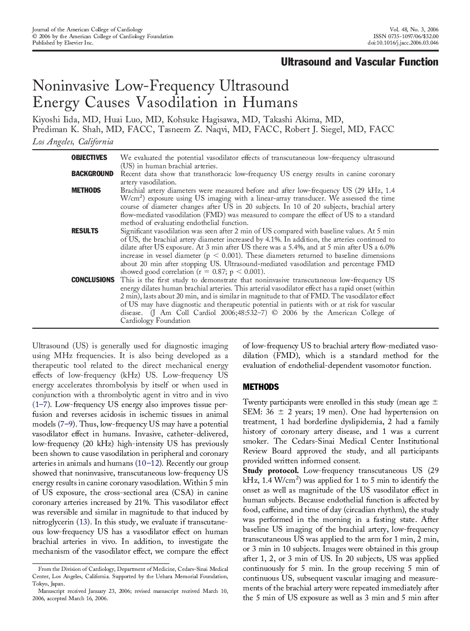
ObjectivesWe evaluated the potential vasodilator effects of transcutaneous low-frequency ultrasound (US) in human brachial arteries.BackgroundRecent data show that transthoracic low-frequency US energy results in canine coronary artery vasodilation.MethodsBrachial artery diameters were measured before and after low-frequency US (29 kHz, 1.4 W/cm2) exposure using US imaging with a linear-array transducer. We assessed the time course of diameter changes after US in 20 subjects. In 10 of 20 subjects, brachial artery flow-mediated vasodilation (FMD) was measured to compare the effect of US to a standard method of evaluating endothelial function.ResultsSignificant vasodilation was seen after 2 min of US compared with baseline values. At 5 min of US, the brachial artery diameter increased by 4.1%. In addition, the arteries continued to dilate after US exposure. At 3 min after US there was a 5.4%, and at 5 min after US a 6.0% increase in vessel diameter (p < 0.001). These diameters returned to baseline dimensions about 20 min after stopping US. Ultrasound-mediated vasodilation and percentage FMD showed good correlation (r = 0.87; p < 0.001).ConclusionsThis is the first study to demonstrate that noninvasive transcutaneous low-frequency US energy dilates human brachial arteries. This arterial vasodilator effect has a rapid onset (within 2 min), lasts about 20 min, and is similar in magnitude to that of FMD. The vasodilator effect of US may have diagnostic and therapeutic potential in patients with or at risk for vascular disease.
