| Article ID | Journal | Published Year | Pages | File Type |
|---|---|---|---|---|
| 5982861 | Journal of the American College of Cardiology | 2014 | 9 Pages |
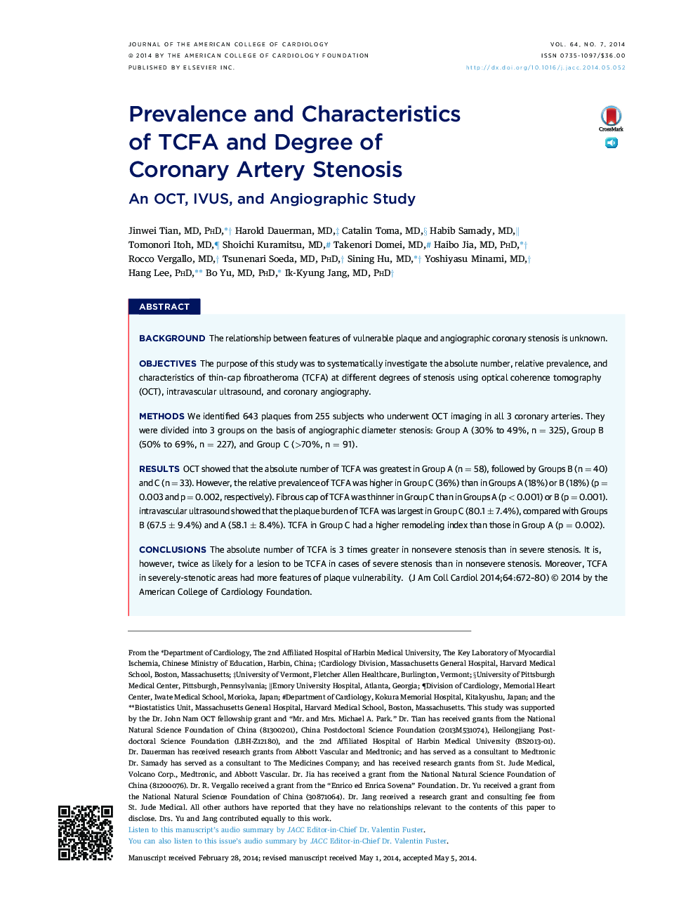
BackgroundThe relationship between features of vulnerable plaque and angiographic coronary stenosis is unknown.ObjectivesThe purpose of this study was to systematically investigate the absolute number, relative prevalence, and characteristics of thin-cap fibroatheroma (TCFA) at different degrees of stenosis using optical coherence tomography (OCT), intravascular ultrasound, and coronary angiography.MethodsWe identified 643 plaques from 255 subjects who underwent OCT imaging in all 3 coronary arteries. They were divided into 3 groups on the basis of angiographic diameter stenosis: Group A (30% to 49%, n = 325), Group B (50% to 69%, n = 227), and Group C (>70%, n = 91).ResultsOCT showed that the absolute number of TCFA was greatest in Group A (n = 58), followed by Groups B (n = 40) and C (n = 33). However, the relative prevalence of TCFA was higher in Group C (36%) than in Groups A (18%) or B (18%) (p = 0.003 and p = 0.002, respectively). Fibrous cap of TCFA was thinner in Group C than in Groups A (p < 0.001) or B (p = 0.001). intravascular ultrasound showed that the plaque burden of TCFA was largest in Group C (80.1 ± 7.4%), compared with Groups B (67.5 ± 9.4%) and A (58.1 ± 8.4%). TCFA in Group C had a higher remodeling index than those in Group A (p = 0.002).ConclusionsThe absolute number of TCFA is 3 times greater in nonsevere stenosis than in severe stenosis. It is, however, twice as likely for a lesion to be TCFA in cases of severe stenosis than in nonsevere stenosis. Moreover, TCFA in severely-stenotic areas had more features of plaque vulnerability.
