| Article ID | Journal | Published Year | Pages | File Type |
|---|---|---|---|---|
| 702899 | Diamond and Related Materials | 2009 | 4 Pages |
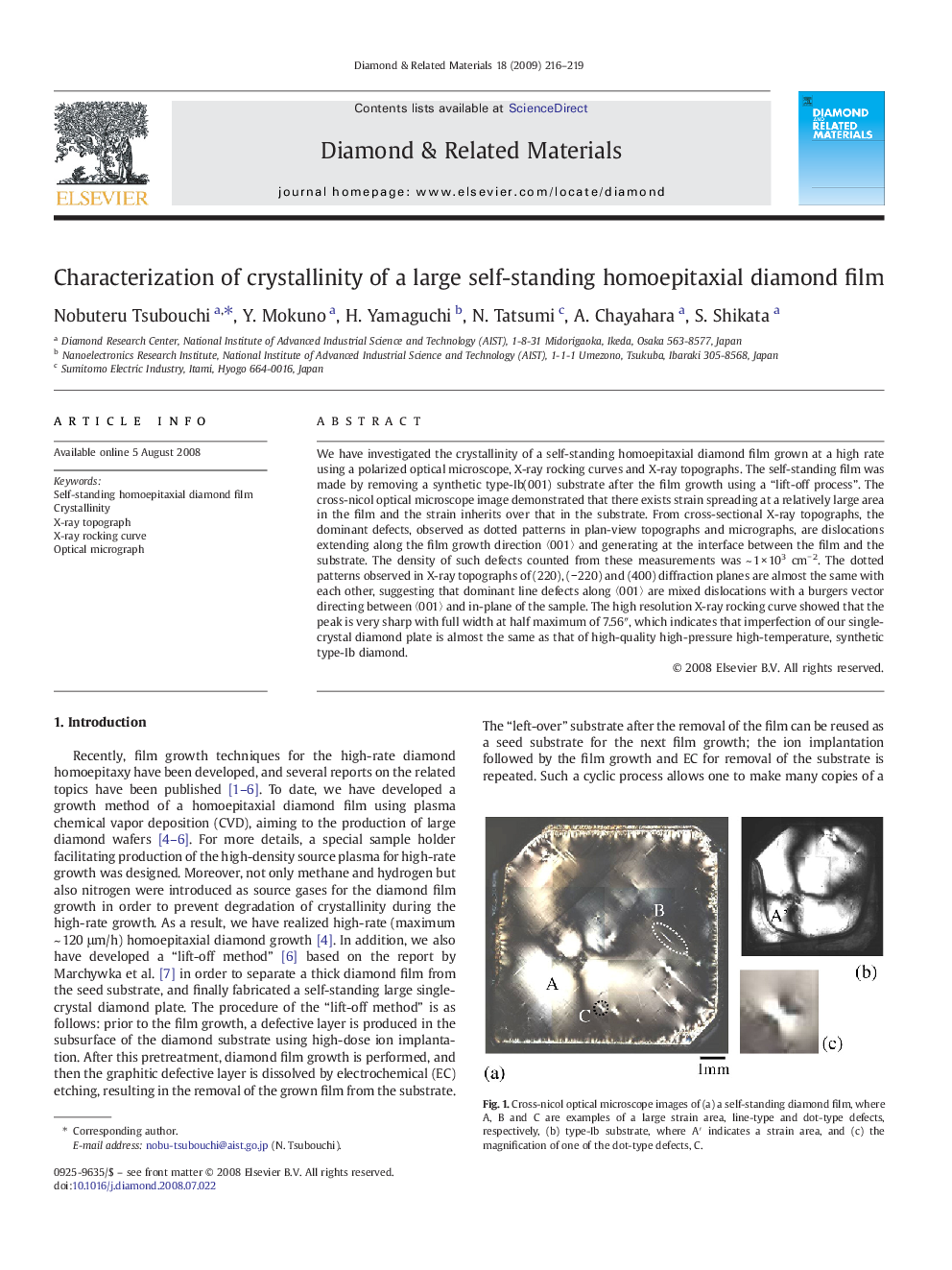
We have investigated the crystallinity of a self-standing homoepitaxial diamond film grown at a high rate using a polarized optical microscope, X-ray rocking curves and X-ray topographs. The self-standing film was made by removing a synthetic type-Ib(001) substrate after the film growth using a “lift-off process”. The cross-nicol optical microscope image demonstrated that there exists strain spreading at a relatively large area in the film and the strain inherits over that in the substrate. From cross-sectional X-ray topographs, the dominant defects, observed as dotted patterns in plan-view topographs and micrographs, are dislocations extending along the film growth direction 〈001〉 and generating at the interface between the film and the substrate. The density of such defects counted from these measurements was ~ 1 × 103 cm− 2. The dotted patterns observed in X-ray topographs of (220), (− 220) and (400) diffraction planes are almost the same with each other, suggesting that dominant line defects along 〈001〉 are mixed dislocations with a burgers vector directing between 〈001〉 and in-plane of the sample. The high resolution X-ray rocking curve showed that the peak is very sharp with full width at half maximum of 7.56″, which indicates that imperfection of our single-crystal diamond plate is almost the same as that of high-quality high-pressure high-temperature, synthetic type-Ib diamond.
