| Article ID | Journal | Published Year | Pages | File Type |
|---|---|---|---|---|
| 1408331 | Journal of Molecular Structure | 2015 | 6 Pages |
•VO2+ doped ZnO–CdS composite synthesized by simple chemical precipitation method.•Average particle size is found to be 17.7 nm.•Spherical like structures were observed in SEM and TEM micrographs.•Ionic bonding is observed between vanadyl ions and its ligands.•Vanadyl ions entered into the host lattice as tetragonally distorted octahedral sites.
VO2+ doped ZnO–CdS composite nanopowder has been synthesized by chemical precipitation method. The prepared sample has been characterized by powder X-ray diffraction (XRD), scanning electron microscopy (SEM), transmission electron microscopy (TEM), FT-IR, photoluminescence (PL), optical absorption and EPR spectroscopy. From XRD pattern, average crystallite size is about 18 nm. SEM and TEM images showed sphere like structures. FT-IR spectrum indicates the presence of fundamental modes of ZnO, CdS and other functional groups. The PL spectrum of VO2+ doped ZnO–CdS composite nanopowder exhibits UV, blue and green emissions. Optical and EPR studies revealed the tetragonal compressed octahedral site symmetry for VO2+ ions. The bonding between VO2+ and its ligands is ionic.
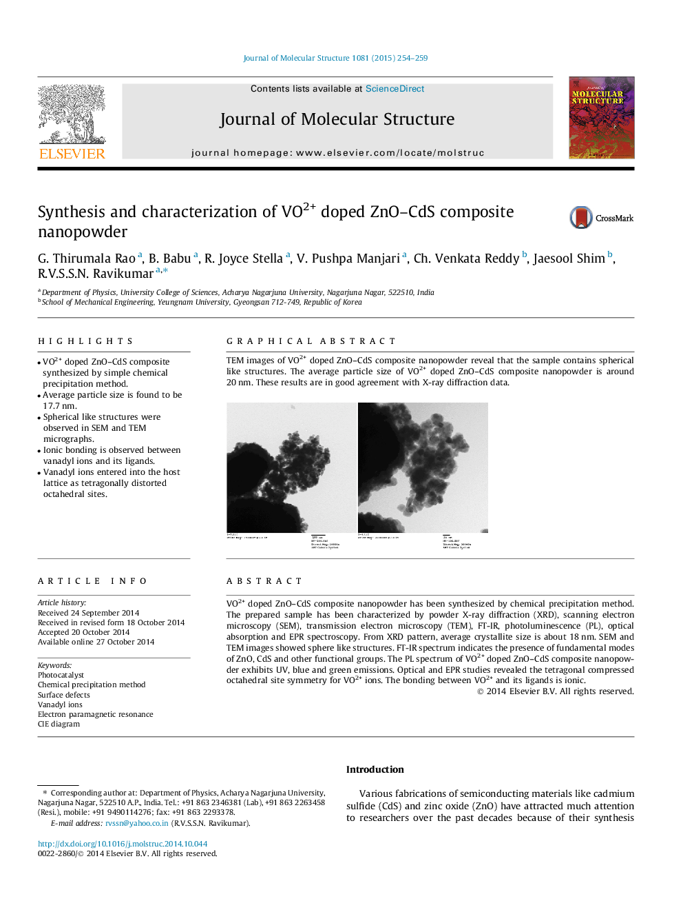
Graphical abstractTEM images of VO2+ doped ZnO–CdS composite nanopowder reveal that the sample contains spherical like structures. The average particle size of VO2+ doped ZnO–CdS composite nanopowder is around 20 nm. These results are in good agreement with X-ray diffraction data.Figure optionsDownload full-size imageDownload as PowerPoint slide
