| کد مقاله | کد نشریه | سال انتشار | مقاله انگلیسی | نسخه تمام متن |
|---|---|---|---|---|
| 9091883 | 1148986 | 2005 | 6 صفحه PDF | دانلود رایگان |
عنوان انگلیسی مقاله ISI
Apport de la spectroscopie RMN à l'évaluation du traumatisme crânien
دانلود مقاله + سفارش ترجمه
دانلود مقاله ISI انگلیسی
رایگان برای ایرانیان
کلمات کلیدی
موضوعات مرتبط
علوم پزشکی و سلامت
پزشکی و دندانپزشکی
بیهوشی و پزشکی درد
پیش نمایش صفحه اول مقاله

چکیده انگلیسی
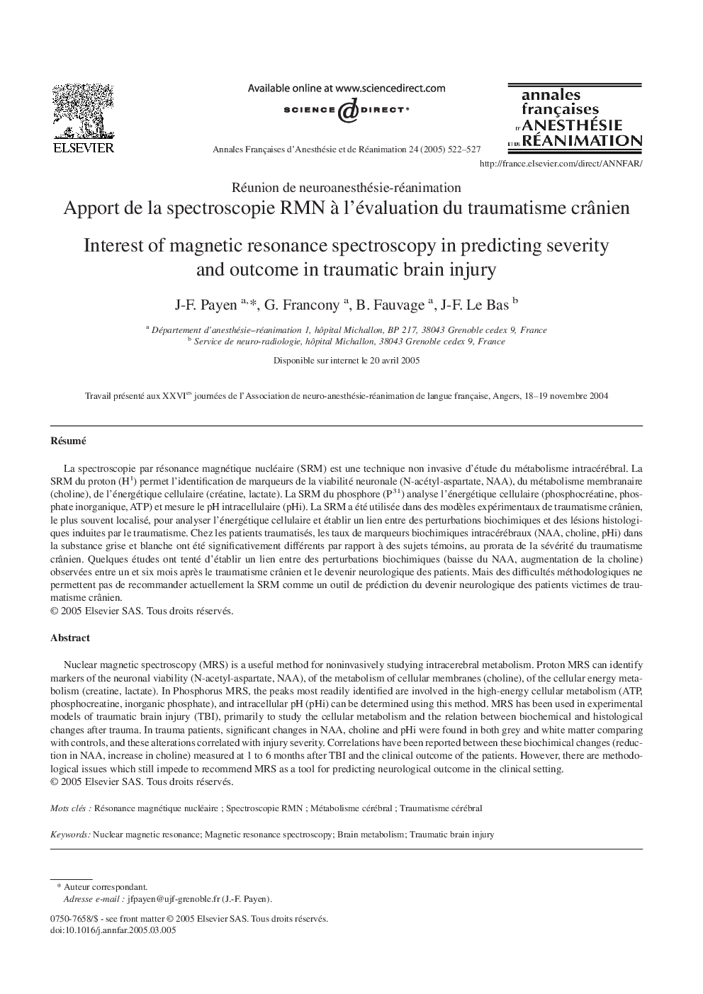
Nuclear magnetic spectroscopy (MRS) is a useful method for noninvasively studying intracerebral metabolism. Proton MRS can identify markers of the neuronal viability (N-acetyl-aspartate, NAA), of the metabolism of cellular membranes (choline), of the cellular energy metabolism (creatine, lactate). In Phosphorus MRS, the peaks most readily identified are involved in the high-energy cellular metabolism (ATP, phosphocreatine, inorganic phosphate), and intracellular pH (pHi) can be determined using this method. MRS has been used in experimental models of traumatic brain injury (TBI), primarily to study the cellular metabolism and the relation between biochemical and histological changes after trauma. In trauma patients, significant changes in NAA, choline and pHi were found in both grey and white matter comparing with controls, and these alterations correlated with injury severity. Correlations have been reported between these biochimical changes (reduction in NAA, increase in choline) measured at 1 to 6Â months after TBI and the clinical outcome of the patients. However, there are methodological issues which still impede to recommend MRS as a tool for predicting neurological outcome in the clinical setting.
ناشر
Database: Elsevier - ScienceDirect (ساینس دایرکت)
Journal: Annales Françaises d'Anesthésie et de Réanimation - Volume 24, Issue 5, May 2005, Pages 522-527
Journal: Annales Françaises d'Anesthésie et de Réanimation - Volume 24, Issue 5, May 2005, Pages 522-527
نویسندگان
J-F. Payen, G. Francony, B. Fauvage, J-F. Le Bas,