| کد مقاله | کد نشریه | سال انتشار | مقاله انگلیسی | نسخه تمام متن |
|---|---|---|---|---|
| 10691564 | 1019601 | 2014 | 11 صفحه PDF | دانلود رایگان |
عنوان انگلیسی مقاله ISI
Monitoring and Staging Abdominal Aortic Aneurysm Disease With Pulse Wave Imaging
ترجمه فارسی عنوان
نظارت و ارزیابی بیماری آئرویسم شکمی شکم با تصویربرداری از موج پالس
دانلود مقاله + سفارش ترجمه
دانلود مقاله ISI انگلیسی
رایگان برای ایرانیان
کلمات کلیدی
High-frequency imagingAbdominal aortic aneurysm - آنوریسمی آئورت شکمیMotion estimation - برآورد حرکتUltrasound imaging - تصویربرداری اولتراسوندSpeckle tracking - ردیابی SpecklePulse wave velocity - سرعت موج پالسFissure - شکافMice - موشNormalized cross-correlation - همبستگی متقابل نرمالRupture - پارگی
موضوعات مرتبط
مهندسی و علوم پایه
فیزیک و نجوم
آکوستیک و فرا صوت
چکیده انگلیسی
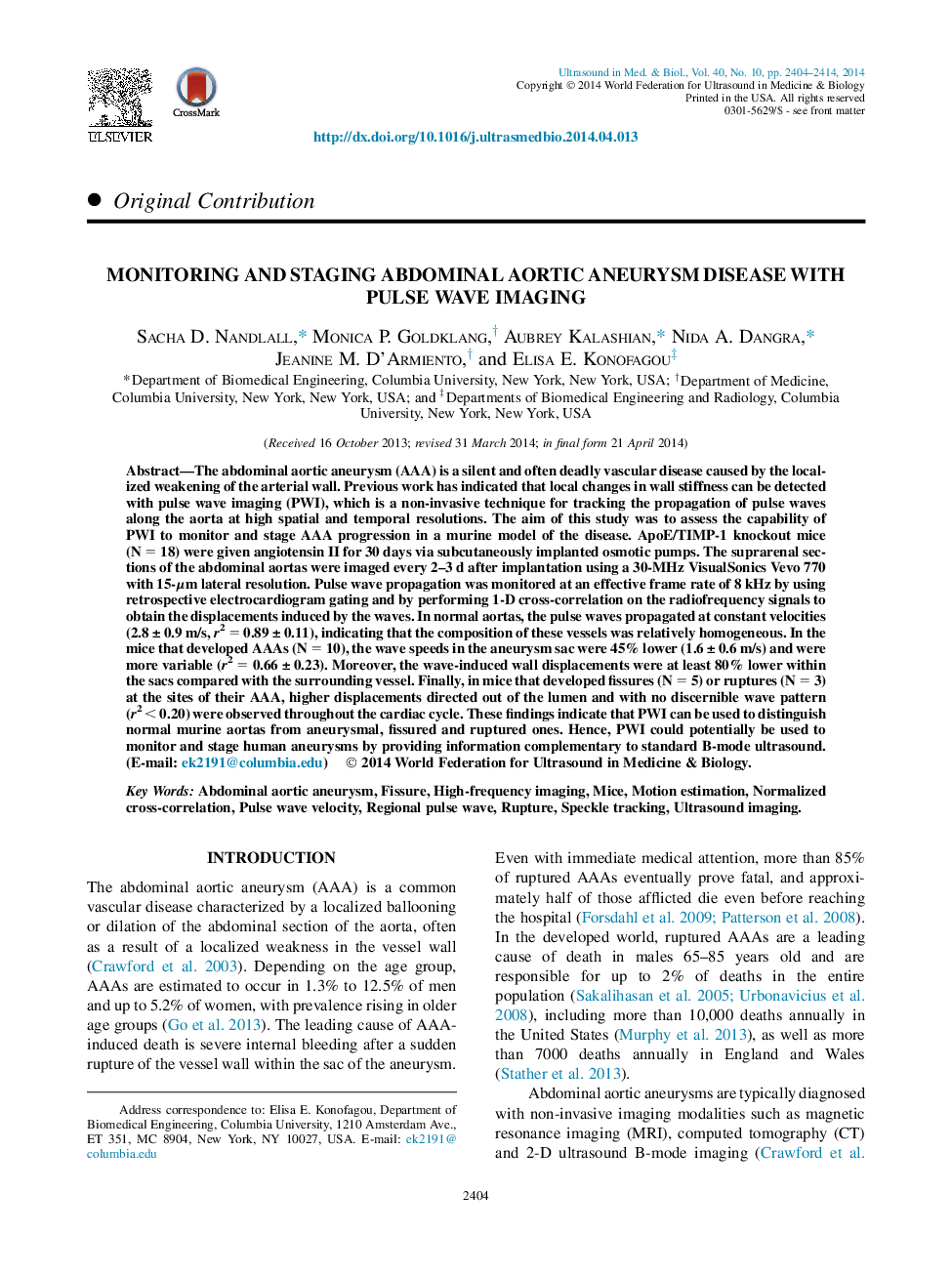
The abdominal aortic aneurysm (AAA) is a silent and often deadly vascular disease caused by the localized weakening of the arterial wall. Previous work has indicated that local changes in wall stiffness can be detected with pulse wave imaging (PWI), which is a non-invasive technique for tracking the propagation of pulse waves along the aorta at high spatial and temporal resolutions. The aim of this study was to assess the capability of PWI to monitor and stage AAA progression in a murine model of the disease. ApoE/TIMP-1 knockout mice (N = 18) were given angiotensin II for 30 days via subcutaneously implanted osmotic pumps. The suprarenal sections of the abdominal aortas were imaged every 2-3 d after implantation using a 30-MHz VisualSonics Vevo 770 with 15-μm lateral resolution. Pulse wave propagation was monitored at an effective frame rate of 8 kHz by using retrospective electrocardiogram gating and by performing 1-D cross-correlation on the radiofrequency signals to obtain the displacements induced by the waves. In normal aortas, the pulse waves propagated at constant velocities (2.8 ± 0.9 m/s, r2 = 0.89 ± 0.11), indicating that the composition of these vessels was relatively homogeneous. In the mice that developed AAAs (N = 10), the wave speeds in the aneurysm sac were 45% lower (1.6 ± 0.6 m/s) and were more variable (r2 = 0.66 ± 0.23). Moreover, the wave-induced wall displacements were at least 80% lower within the sacs compared with the surrounding vessel. Finally, in mice that developed fissures (N = 5) or ruptures (N = 3) at the sites of their AAA, higher displacements directed out of the lumen and with no discernible wave pattern (r2 < 0.20) were observed throughout the cardiac cycle. These findings indicate that PWI can be used to distinguish normal murine aortas from aneurysmal, fissured and ruptured ones. Hence, PWI could potentially be used to monitor and stage human aneurysms by providing information complementary to standard B-mode ultrasound.
ناشر
Database: Elsevier - ScienceDirect (ساینس دایرکت)
Journal: Ultrasound in Medicine & Biology - Volume 40, Issue 10, October 2014, Pages 2404-2414
Journal: Ultrasound in Medicine & Biology - Volume 40, Issue 10, October 2014, Pages 2404-2414
نویسندگان
Sacha D. Nandlall, Monica P. Goldklang, Aubrey Kalashian, Nida A. Dangra, Jeanine M. D'Armiento, Elisa E. Konofagou,
