| کد مقاله | کد نشریه | سال انتشار | مقاله انگلیسی | نسخه تمام متن |
|---|---|---|---|---|
| 1760276 | 1019587 | 2015 | 10 صفحه PDF | دانلود رایگان |
عنوان انگلیسی مقاله ISI
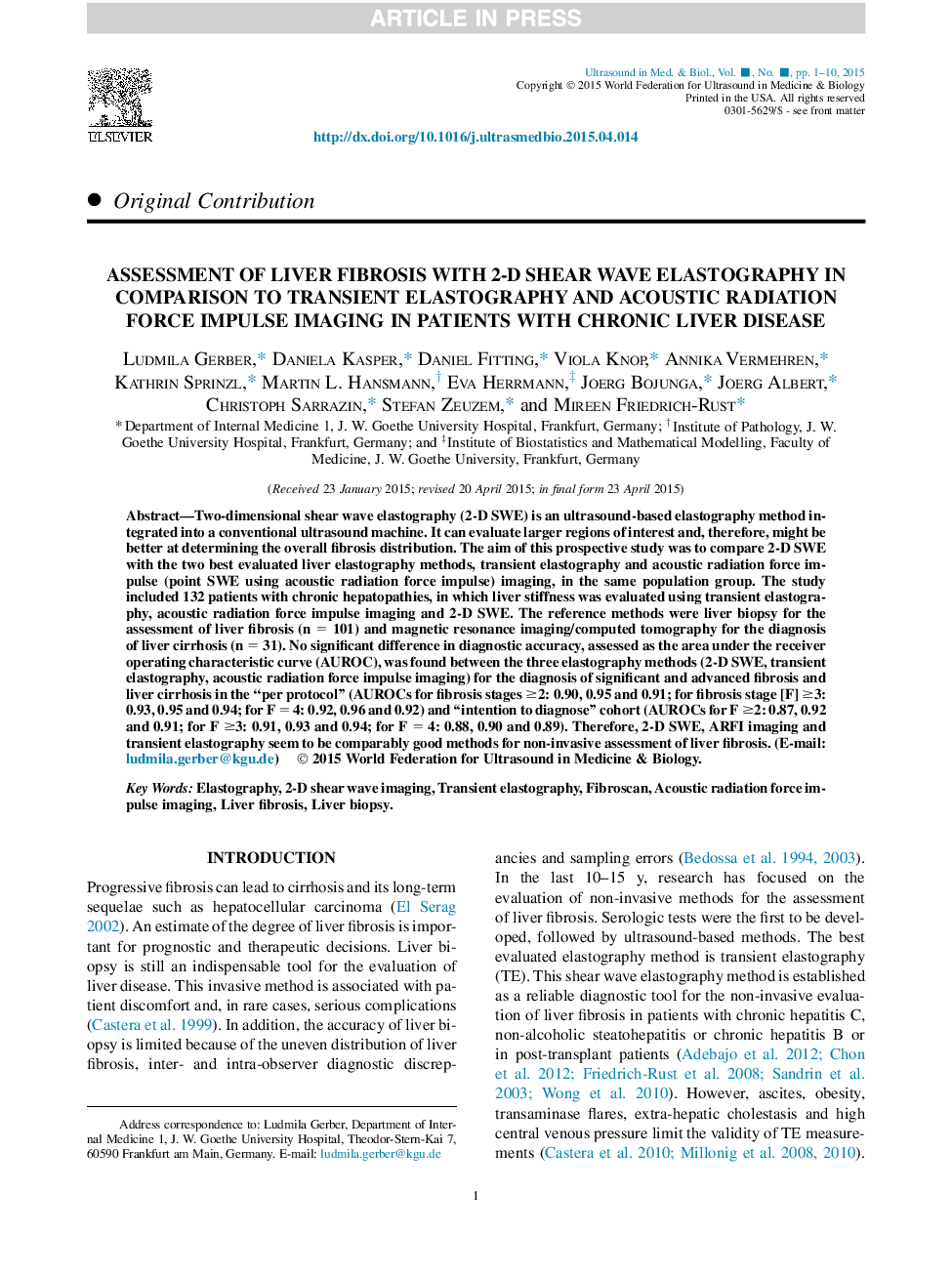
Assessment of Liver Fibrosis with 2-D Shear Wave Elastography in Comparison to Transient Elastography and Acoustic Radiation Force Impulse Imaging in Patients with Chronic Liver Disease
دانلود مقاله + سفارش ترجمه
دانلود مقاله ISI انگلیسی
رایگان برای ایرانیان
کلمات کلیدی
موضوعات مرتبط
مهندسی و علوم پایه
فیزیک و نجوم
آکوستیک و فرا صوت
پیش نمایش صفحه اول مقاله

چکیده انگلیسی
Two-dimensional shear wave elastography (2-D SWE) is an ultrasound-based elastography method integrated into a conventional ultrasound machine. It can evaluate larger regions of interest and, therefore, might be better at determining the overall fibrosis distribution. The aim of this prospective study was to compare 2-D SWE with the two best evaluated liver elastography methods, transient elastography and acoustic radiation force impulse (point SWE using acoustic radiation force impulse) imaging, in the same population group. The study included 132 patients with chronic hepatopathies, in which liver stiffness was evaluated using transient elastography, acoustic radiation force impulse imaging and 2-D SWE. The reference methods were liver biopsy for the assessment of liver fibrosis (n = 101) and magnetic resonance imaging/computed tomography for the diagnosis of liver cirrhosis (n = 31). No significant difference in diagnostic accuracy, assessed as the area under the receiver operating characteristic curve (AUROC), was found between the three elastography methods (2-D SWE, transient elastography, acoustic radiation force impulse imaging) for the diagnosis of significant and advanced fibrosis and liver cirrhosis in the “per protocol” (AUROCs for fibrosis stages â¥2: 0.90, 0.95 and 0.91; for fibrosis stage [F] â¥3: 0.93, 0.95 and 0.94; for F = 4: 0.92, 0.96 and 0.92) and “intention to diagnose” cohort (AUROCs for F â¥2: 0.87, 0.92 and 0.91; for F â¥3: 0.91, 0.93 and 0.94; for F = 4: 0.88, 0.90 and 0.89). Therefore, 2-D SWE, ARFI imaging and transient elastography seem to be comparably good methods for non-invasive assessment of liver fibrosis.
ناشر
Database: Elsevier - ScienceDirect (ساینس دایرکت)
Journal: Ultrasound in Medicine & Biology - Volume 41, Issue 9, September 2015, Pages 2350-2359
Journal: Ultrasound in Medicine & Biology - Volume 41, Issue 9, September 2015, Pages 2350-2359
نویسندگان
Ludmila Gerber, Daniela Kasper, Daniel Fitting, Viola Knop, Annika Vermehren, Kathrin Sprinzl, Martin L. Hansmann, Eva Herrmann, Joerg Bojunga, Joerg Albert, Christoph Sarrazin, Stefan Zeuzem, Mireen Friedrich-Rust,