| کد مقاله | کد نشریه | سال انتشار | مقاله انگلیسی | نسخه تمام متن |
|---|---|---|---|---|
| 8130809 | 1523230 | 2018 | 7 صفحه PDF | دانلود رایگان |
عنوان انگلیسی مقاله ISI
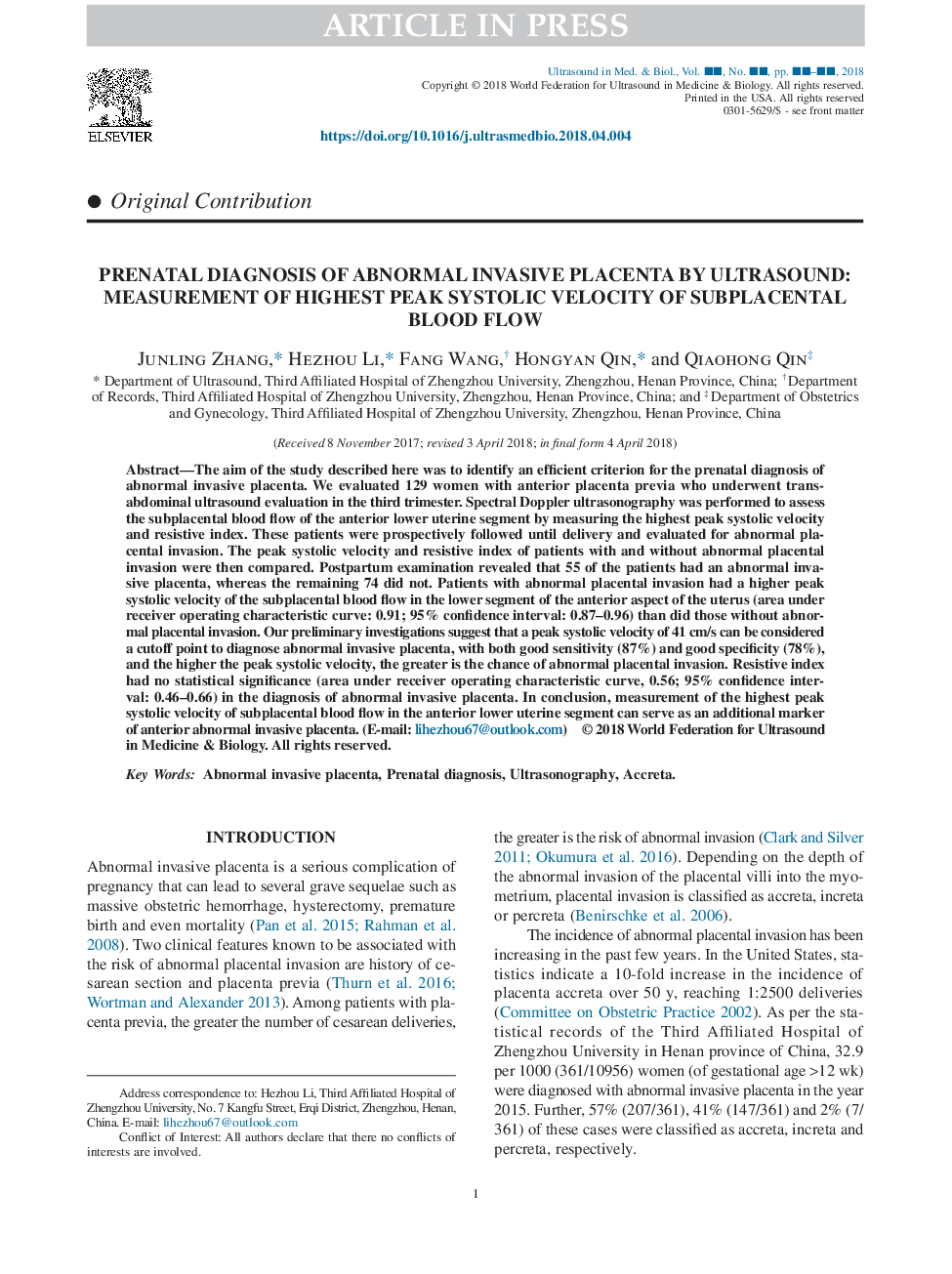
Prenatal Diagnosis of Abnormal Invasive Placenta by Ultrasound: Measurement of Highest Peak Systolic Velocity of Subplacental Blood Flow
ترجمه فارسی عنوان
تشخیص پیش از قاعدگی از انسداد غیر عادی انسدادی توسط سونوگرافی: اندازه گیری سرعت بالاتر سی تی اسکن پیک خون جریان زیر پوستی
دانلود مقاله + سفارش ترجمه
دانلود مقاله ISI انگلیسی
رایگان برای ایرانیان
کلمات کلیدی
جفت نابالغ تهاجمی، تشخیص قبل از تولد، سونوگرافی، آکریتا،
موضوعات مرتبط
مهندسی و علوم پایه
فیزیک و نجوم
آکوستیک و فرا صوت
چکیده انگلیسی
The aim of the study described here was to identify an efficient criterion for the prenatal diagnosis of abnormal invasive placenta. We evaluated 129 women with anterior placenta previa who underwent trans-abdominal ultrasound evaluation in the third trimester. Spectral Doppler ultrasonography was performed to assess the subplacental blood flow of the anterior lower uterine segment by measuring the highest peak systolic velocity and resistive index. These patients were prospectively followed until delivery and evaluated for abnormal placental invasion. The peak systolic velocity and resistive index of patients with and without abnormal placental invasion were then compared. Postpartum examination revealed that 55 of the patients had an abnormal invasive placenta, whereas the remaining 74 did not. Patients with abnormal placental invasion had a higher peak systolic velocity of the subplacental blood flow in the lower segment of the anterior aspect of the uterus (area under receiver operating characteristic curve: 0.91; 95% confidence interval: 0.87-0.96) than did those without abnormal placental invasion. Our preliminary investigations suggest that a peak systolic velocity of 41âcm/s can be considered a cutoff point to diagnose abnormal invasive placenta, with both good sensitivity (87%) and good specificity (78%), and the higher the peak systolic velocity, the greater is the chance of abnormal placental invasion. Resistive index had no statistical significance (area under receiver operating characteristic curve, 0.56; 95% confidence interval: 0.46-0.66) in the diagnosis of abnormal invasive placenta. In conclusion, measurement of the highest peak systolic velocity of subplacental blood flow in the anterior lower uterine segment can serve as an additional marker of anterior abnormal invasive placenta.
ناشر
Database: Elsevier - ScienceDirect (ساینس دایرکت)
Journal: Ultrasound in Medicine & Biology - Volume 44, Issue 8, August 2018, Pages 1672-1678
Journal: Ultrasound in Medicine & Biology - Volume 44, Issue 8, August 2018, Pages 1672-1678
نویسندگان
Junling Zhang, Hezhou Li, Fang Wang, Hongyan Qin, Qiaohong Qin,
