| کد مقاله | کد نشریه | سال انتشار | مقاله انگلیسی | نسخه تمام متن |
|---|---|---|---|---|
| 9390516 | 1282819 | 2005 | 18 صفحه PDF | دانلود رایگان |
عنوان انگلیسی مقاله ISI
Imagerie de la neurofibromatose de type 1
دانلود مقاله + سفارش ترجمه
دانلود مقاله ISI انگلیسی
رایگان برای ایرانیان
کلمات کلیدی
موضوعات مرتبط
علوم پزشکی و سلامت
پزشکی و دندانپزشکی
رادیولوژی و تصویربرداری
پیش نمایش صفحه اول مقاله

چکیده انگلیسی
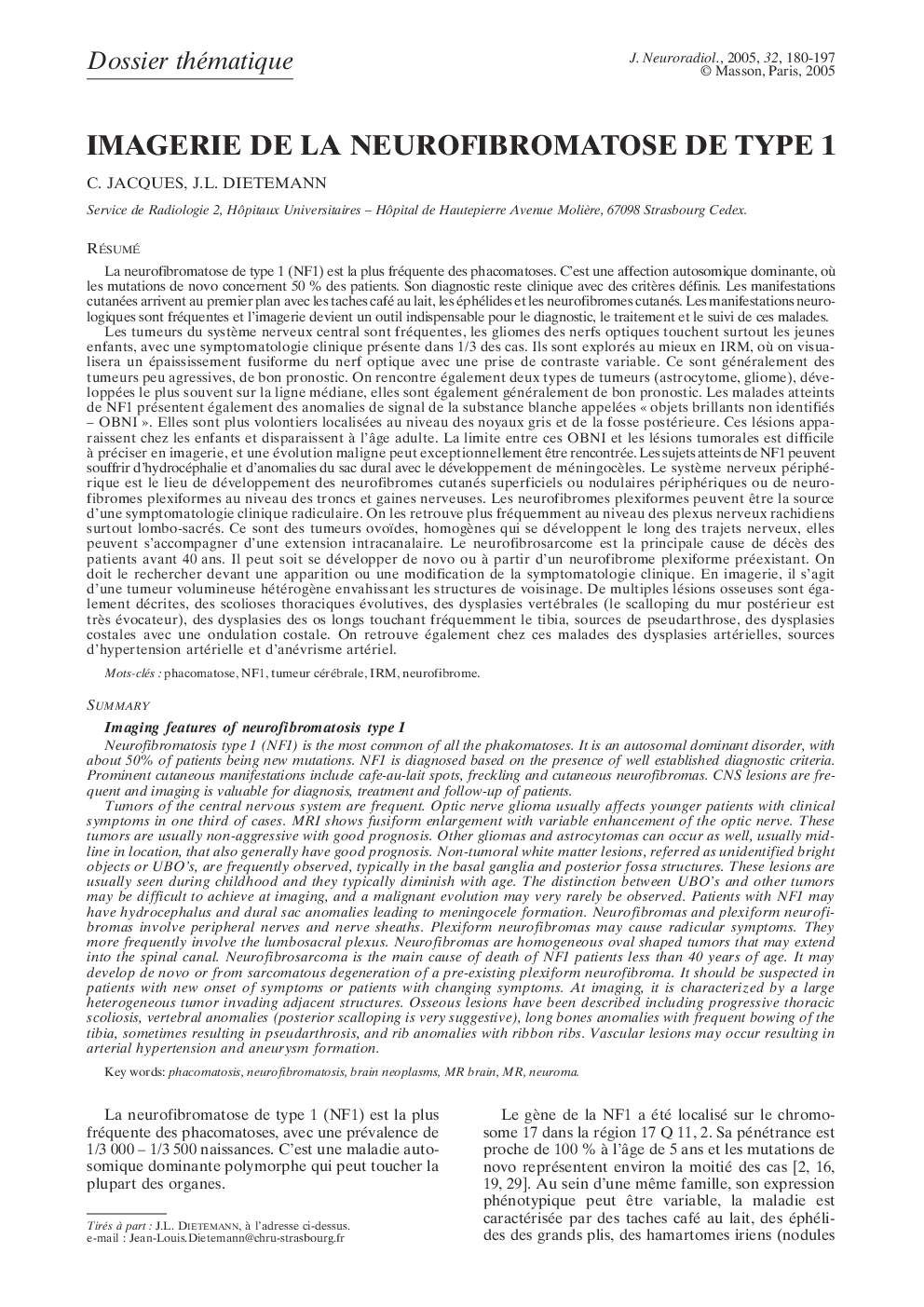
Tumors of the central nervous system are frequent. Optic nerve glioma usually affects younger patients with clinical symptoms in one third of cases. MRI shows fusiform enlargement with variable enhancement of the optic nerve. These tumors are usually non-aggressive with good prognosis. Other gliomas and astrocytomas can occur as well, usually midline in location, that also generally have good prognosis. Non-tumoral white matter lesions, referred as unidentified bright objects or UBO's, are frequently observed, typically in the basal ganglia and posterior fossa structures. These lesions are usually seen during childhood and they typically diminish with age. The distinction between UBO's and other tumors may be difficult to achieve at imaging, and a malignant evolution may very rarely be observed. Patients with NF1 may have hydrocephalus and dural sac anomalies leading to meningocele formation. Neurofibromas and plexiform neurofibromas involve peripheral nerves and nerve sheaths. Plexiform neurofibromas may cause radicular symptoms. They more frequently involve the lumbosacral plexus. Neurofibromas are homogeneous oval shaped tumors that may extend into the spinal canal. Neurofibrosarcoma is the main cause of death of NF1 patients less than 40 years of age. It may develop de novo or from sarcomatous degeneration of a pre-existing plexiform neurofibroma. It should be suspected in patients with new onset of symptoms or patients with changing symptoms. At imaging, it is characterized by a large heterogeneous tumor invading adjacent structures. Osseous lesions have been described including progressive thoracic scoliosis, vertebral anomalies (posterior scalloping is very suggestive), long bones anomalies with frequent bowing of the tibia, sometimes resulting in pseudarthrosis, and rib anomalies with ribbon ribs. Vascular lesions may occur resulting in arterial hypertension and aneurysm formation.
ناشر
Database: Elsevier - ScienceDirect (ساینس دایرکت)
Journal: Journal of Neuroradiology - Volume 32, Issue 3, June 2005, Pages 180-197
Journal: Journal of Neuroradiology - Volume 32, Issue 3, June 2005, Pages 180-197
نویسندگان
C. Jacques, J.L. Dietemann,