| کد مقاله | کد نشریه | سال انتشار | مقاله انگلیسی | نسخه تمام متن |
|---|---|---|---|---|
| 5491418 | 1525006 | 2017 | 12 صفحه PDF | دانلود رایگان |
عنوان انگلیسی مقاله ISI
Identifying non-toxic doses of manganese for manganese-enhanced magnetic resonance imaging to map brain areas activated by operant behavior in trained rats
ترجمه فارسی عنوان
شناسایی دوزهای غیر سمی منگنز برای تصویربرداری رزونانس مغناطیسی با منگنز برای نقشه برداری مناطق مغز فعال شده توسط رفتار اپراتور در موشهای آموزش دیده
دانلود مقاله + سفارش ترجمه
دانلود مقاله ISI انگلیسی
رایگان برای ایرانیان
کلمات کلیدی
ALTPTAMEAaHIVPMRSARRFPRHVPLENTRSGICJectorhinal cortexretrosplenial agranular cortexECTCPUAcb - ACBAST - آسپارتات ترانس آمینازAspartate aminotransferase - آسپارتات ترانس آمیناز یا AST Alanine aminotransferase - آلانین آمینوترانسفرازAmygdala - آمیگدال، بادامهAMY - امیPir - بیش از حدsubstantia nigra - توده سیاهOlfactory tubercle - تومور رحمیislands of Calleja - جزایر Callejaretrorubral field - زمینه بازتولیدSubiculum - زیرکیولومTemporal association cortex - قشر ارتباط موقتیprimary somatosensory cortex - قشر اسموتیسنسوری اولیهEntorhinal cortex - قشر انتورینالprimary motor cortex - قشر حرکتی اولیهsecondary motor cortex - قشر حرکتی ثانویهretrosplenial granular cortex - قشر دانه گربه ای مجزاRetrosplenial cortex - قشر رتروپلاسمیAgranular insular cortex - قشر ساحلی AgranularGranular insular cortex - قشر ساقه غدهdysgranular insular cortex - قشر ساکن غده تیروئیدAuditory Cortex - قشر شنواییperirhinal cortex - قشر پریحینالpiriform cortex - قشر پریکومSecondary somatosensory cortex - قشر کمخونی ثانویهHIP - مفصل ران یا مفصل هیپamygdalohippocampal area - منطقه amygdalohippocampalNucleus accumbens - هسته accumbensVentral posteromedial thalamic nucleus - هسته تالاموم پس از معدهventral posterolateral thalamic nucleus - هسته تالامیک پست سلولی شکمیHypothalamus - هیپوتالاموسHippocampus - هیپوکامپ TEA - چایparietal association cortex - کورتکس انجمن مشترکcingulate cortex - کورتکس کانگولتPit - گودالGlobus pallidus - گوی رنگ پریده، گلوبوس پالیدوس
موضوعات مرتبط
مهندسی و علوم پایه
فیزیک و نجوم
فیزیک ماده چگال
چکیده انگلیسی
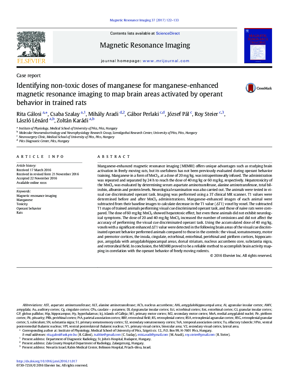
Manganese-enhanced magnetic resonance imaging (MEMRI) offers unique advantages such as studying brain activation in freely moving rats, but its usefulness has not been previously evaluated during operant behavior training. Manganese in a form of MnCl2, at a dose of 20Â mg/kg, was intraperitoneally infused. The administration was repeated and separated by 24Â h to reach the dose of 40Â mg/kg or 60Â mg/kg, respectively. Hepatotoxicity of the MnCl2 was evaluated by determining serum aspartate aminotransferase, alanine aminotransferase, total bilirubin, albumin and protein levels. Neurological examination was also carried out. The animals were tested in visual cue discriminated operant task. Imaging was performed using a 3T clinical MR scanner. T1 values were determined before and after MnCl2 administrations. Manganese-enhanced images of each animal were subtracted from their baseline images to calculate decrease in the T1 value (ÎT1) voxel by voxel. The subtracted T1 maps of trained animals performing visual cue discriminated operant task, and those of naive rats were compared. The dose of 60Â mg/kg MnCl2 showed hepatotoxic effect, but even these animals did not exhibit neurological symptoms. The dose of 20 and 40Â mg/kg MnCl2 increased the number of omissions and did not affect the accuracy of performing the visual cue discriminated operant task. Using the accumulated dose of 40Â mg/kg, voxels with a significant enhanced ÎT1 value were detected in the following brain areas of the visual cue discriminated operant behavior performed animals compared to those in the controls: the visual, somatosensory, motor and premotor cortices, the insula, cingulate, ectorhinal, entorhinal, perirhinal and piriform cortices, hippocampus, amygdala with amygdalohippocampal areas, dorsal striatum, nucleus accumbens core, substantia nigra, and retrorubral field. In conclusion, the MEMRI proved to be a reliable method to accomplish brain activity mapping in correlation with the operant behavior of freely moving rodents.
ناشر
Database: Elsevier - ScienceDirect (ساینس دایرکت)
Journal: Magnetic Resonance Imaging - Volume 37, April 2017, Pages 122-133
Journal: Magnetic Resonance Imaging - Volume 37, April 2017, Pages 122-133
نویسندگان
Rita Gálosi, Csaba Szalay, Mihály Aradi, Gábor Perlaki, József Pál, Roy Steier, László Lénárd, Zoltán Karádi,
