| کد مقاله | کد نشریه | سال انتشار | مقاله انگلیسی | نسخه تمام متن |
|---|---|---|---|---|
| 1914812 | 1535175 | 2009 | 7 صفحه PDF | دانلود رایگان |
عنوان انگلیسی مقاله ISI
Radiological features of craniopharyngiomas located in the posterior fossa
دانلود مقاله + سفارش ترجمه
دانلود مقاله ISI انگلیسی
رایگان برای ایرانیان
کلمات کلیدی
موضوعات مرتبط
علوم زیستی و بیوفناوری
بیوشیمی، ژنتیک و زیست شناسی مولکولی
سالمندی
پیش نمایش صفحه اول مقاله

چکیده انگلیسی
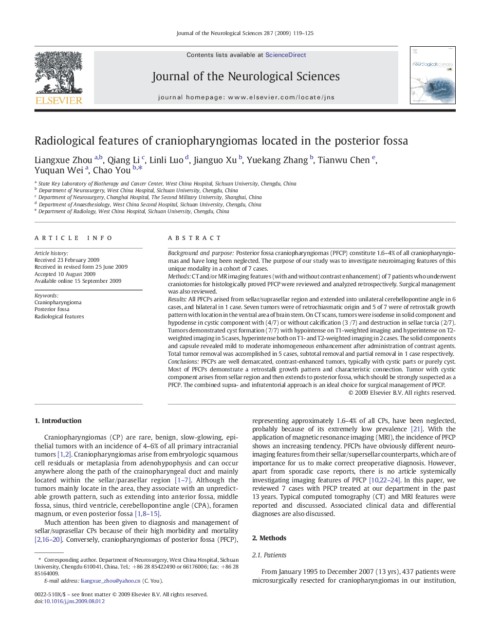
PFCPs are well demarcated, contrast-enhanced tumors, typically with cystic parts or purely cyst. Most of PFCPs demonstrate a retrostalk growth pattern and characteristic connection. Tumor with cystic component arises from sellar region and then extends to posterior fossa, which should be strongly suspected as a PFCP. The combined supra- and infratentorial approach is an ideal choice for surgical management of PFCP.
ناشر
Database: Elsevier - ScienceDirect (ساینس دایرکت)
Journal: Journal of the Neurological Sciences - Volume 287, Issues 1â2, 15 December 2009, Pages 119-125
Journal: Journal of the Neurological Sciences - Volume 287, Issues 1â2, 15 December 2009, Pages 119-125
نویسندگان
Liangxue Zhou, Qiang Li, Linli Luo, Jianguo Xu, Yuekang Zhang, Tianwu Chen, Yuquan Wei, Chao You,