| کد مقاله | کد نشریه | سال انتشار | مقاله انگلیسی | نسخه تمام متن |
|---|---|---|---|---|
| 2058785 | 1543968 | 2015 | 5 صفحه PDF | دانلود رایگان |

It is important to accurately estimate the electromyogram (EMG)/force relationship of triceps surae (TS) muscle for detecting strength deficit of tibalis anterior (TA) muscle. In literature, the protocol for recording EMG and force of dorsiflexion have been described, and the necessity for immobilizing the ankle has been explained. However, there is a significant variability of the results among researchers even though they report the fixation of the ankle.We have determined that toe extension can cause significant variation in the dorsiflexion force and EMG of TS and this can occur despite following the current guidelines which require immobilizing the ankle. The results also show that there was a large increase in the variability of the force and the RMS of EMG of TS when the toes were not strapped compared with when they were strapped. Thus, with the current guidelines, where there are no instructions regarding the necessity of strapping the toes, the EMG/force relationship of TS could be incorrect and give an inaccurate assessment of the dorsiflexor TA strength.In summary,
• Current methodology to estimate the dorsiflexor TA strength with respect to the TS activity, emphasizing on ankle immobilization is insufficient to prevent large variability in the measurements.
• Toe extension during dorsiflexion was found to be one source of variability in estimating the TA strength.
• It is recommended that guidelines for recording force and EMG from TA and TS muscles should require the strapping of the toes along with the need for immobilizing the ankle.
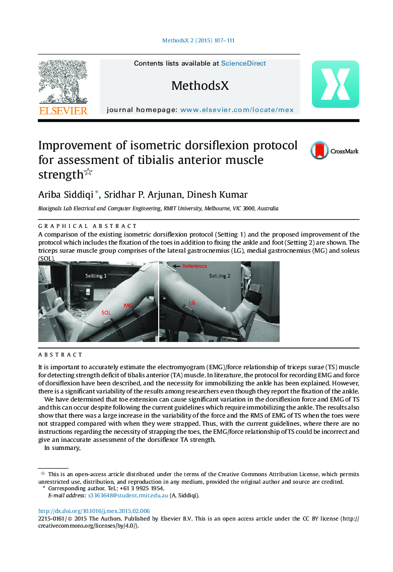
A comparison of the existing isometric dorsiflexion protocol (Setting 1) and the proposed improvement of the protocol which includes the fixation of the toes in addition to fixing the ankle and foot (Setting 2) are shown. The triceps surae muscle group comprises of the lateral gastrocnemius (LG), medial gastrocnemius (MG) and soleus (SOL).Figure optionsDownload as PowerPoint slide
Journal: MethodsX - Volume 2, 2015, Pages 107–111