| کد مقاله | کد نشریه | سال انتشار | مقاله انگلیسی | نسخه تمام متن |
|---|---|---|---|---|
| 2396126 | 1101600 | 2008 | 8 صفحه PDF | دانلود رایگان |

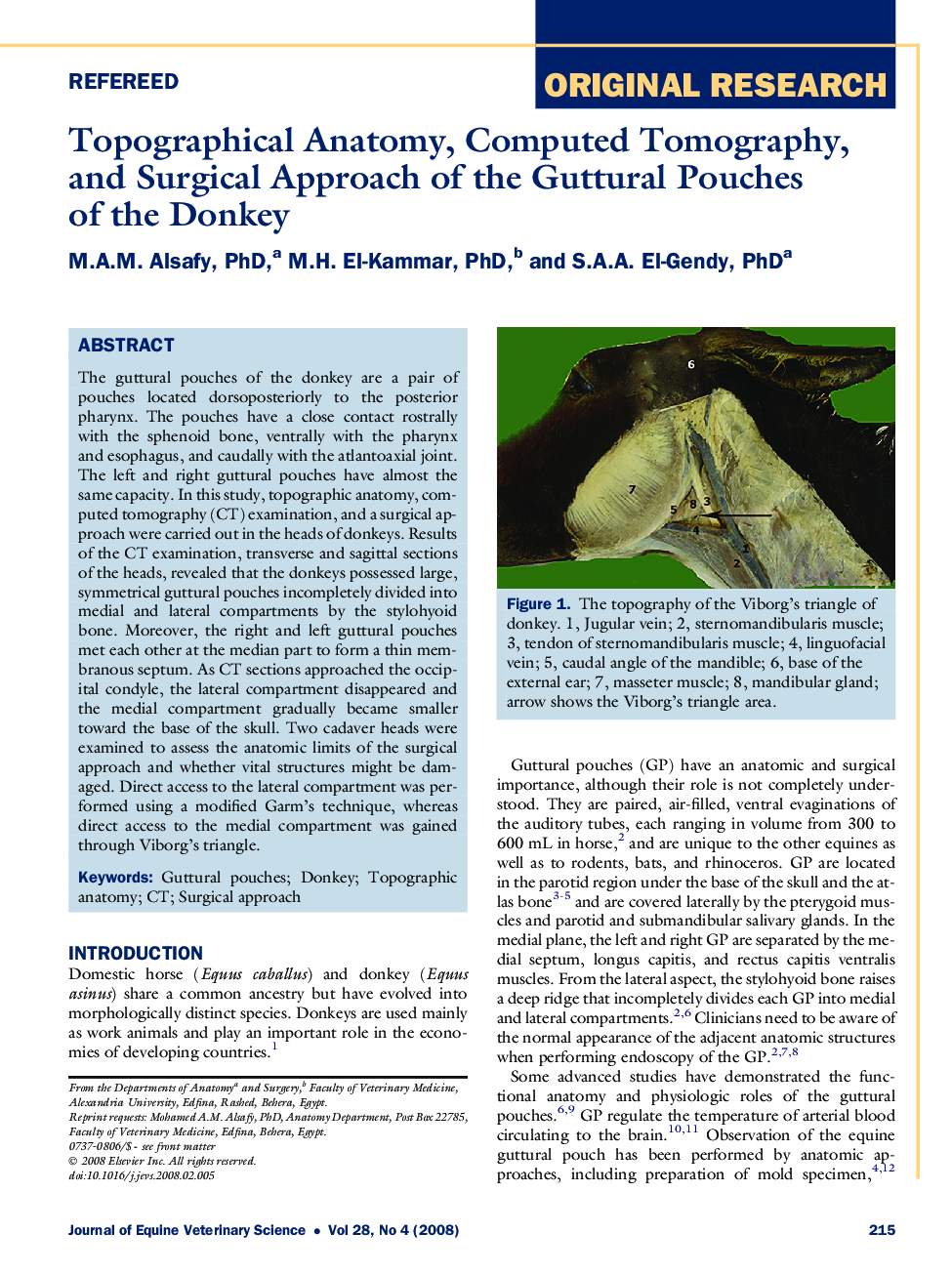
The guttural pouches of the donkey are a pair of pouches located dorsoposteriorly to the posterior pharynx. The pouches have a close contact rostrally with the sphenoid bone, ventrally with the pharynx and esophagus, and caudally with the atlantoaxial joint. The left and right guttural pouches have almost the same capacity. In this study, topographic anatomy, computed tomography (CT) examination, and a surgical approach were carried out in the heads of donkeys. Results of the CT examination, transverse and sagittal sections of the heads, revealed that the donkeys possessed large, symmetrical guttural pouches incompletely divided into medial and lateral compartments by the stylohyoid bone. Moreover, the right and left guttural pouches met each other at the median part to form a thin membranous septum. As CT sections approached the occipital condyle, the lateral compartment disappeared and the medial compartment gradually became smaller toward the base of the skull. Two cadaver heads were examined to assess the anatomic limits of the surgical approach and whether vital structures might be damaged. Direct access to the lateral compartment was performed using a modified Garm's technique, whereas direct access to the medial compartment was gained through Viborg's triangle.
Journal: Journal of Equine Veterinary Science - Volume 28, Issue 4, April 2008, Pages 215–222