| کد مقاله | کد نشریه | سال انتشار | مقاله انگلیسی | نسخه تمام متن |
|---|---|---|---|---|
| 5586565 | 1568637 | 2017 | 20 صفحه PDF | دانلود رایگان |
عنوان انگلیسی مقاله ISI
Three-dimensional reconstruction of the embryonic pancreas in the grass snake Natrix natrix L. (Lepidosauria, Serpentes) based on histological studies
دانلود مقاله + سفارش ترجمه
دانلود مقاله ISI انگلیسی
رایگان برای ایرانیان
موضوعات مرتبط
علوم زیستی و بیوفناوری
علوم کشاورزی و بیولوژیک
علوم دامی و جانورشناسی
پیش نمایش صفحه اول مقاله

چکیده انگلیسی
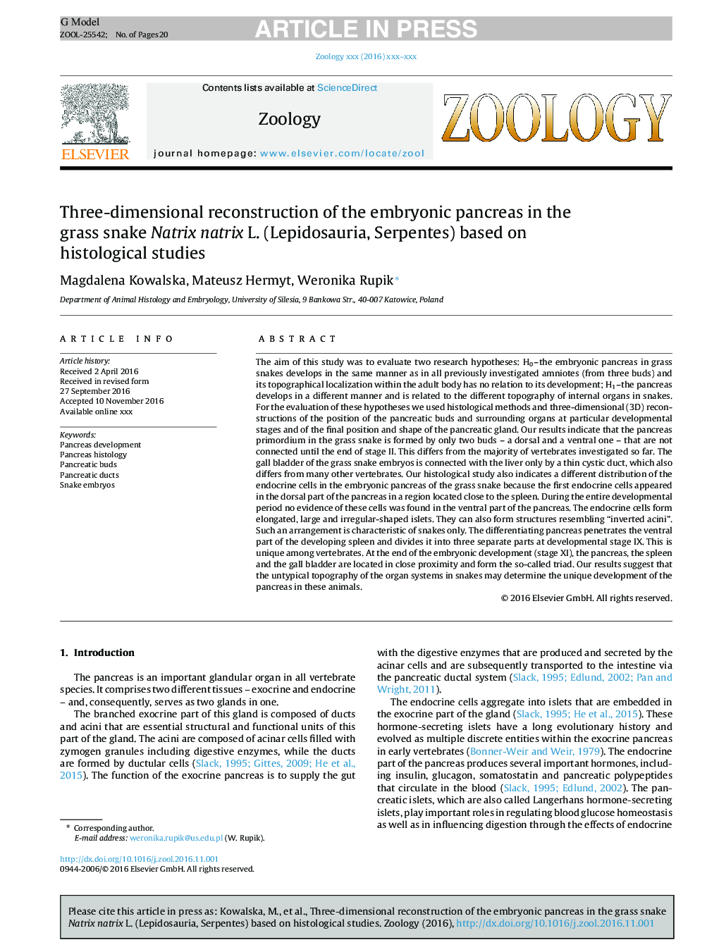
The aim of this study was to evaluate two research hypotheses: H0-the embryonic pancreas in grass snakes develops in the same manner as in all previously investigated amniotes (from three buds) and its topographical localization within the adult body has no relation to its development; H1-the pancreas develops in a different manner and is related to the different topography of internal organs in snakes. For the evaluation of these hypotheses we used histological methods and three-dimensional (3D) reconstructions of the position of the pancreatic buds and surrounding organs at particular developmental stages and of the final position and shape of the pancreatic gland. Our results indicate that the pancreas primordium in the grass snake is formed by only two buds - a dorsal and a ventral one - that are not connected until the end of stage II. This differs from the majority of vertebrates investigated so far. The gall bladder of the grass snake embryos is connected with the liver only by a thin cystic duct, which also differs from many other vertebrates. Our histological study also indicates a different distribution of the endocrine cells in the embryonic pancreas of the grass snake because the first endocrine cells appeared in the dorsal part of the pancreas in a region located close to the spleen. During the entire developmental period no evidence of these cells was found in the ventral part of the pancreas. The endocrine cells form elongated, large and irregular-shaped islets. They can also form structures resembling “inverted acini”. Such an arrangement is characteristic of snakes only. The differentiating pancreas penetrates the ventral part of the developing spleen and divides it into three separate parts at developmental stage IX. This is unique among vertebrates. At the end of the embryonic development (stage XI), the pancreas, the spleen and the gall bladder are located in close proximity and form the so-called triad. Our results suggest that the untypical topography of the organ systems in snakes may determine the unique development of the pancreas in these animals.
ناشر
Database: Elsevier - ScienceDirect (ساینس دایرکت)
Journal: Zoology - Volume 121, April 2017, Pages 91-110
Journal: Zoology - Volume 121, April 2017, Pages 91-110
نویسندگان
Magdalena Kowalska, Mateusz Hermyt, Weronika Rupik,