| کد مقاله | کد نشریه | سال انتشار | مقاله انگلیسی | نسخه تمام متن |
|---|---|---|---|---|
| 606673 | 1454538 | 2015 | 8 صفحه PDF | دانلود رایگان |

• MnO NPs were prepared by a thermal decomposition method.
• Water dispersible MnO NPs were obtained by carboxyl silane modification.
• PEG-Cy5.5 conjugated MnO NPs were fabricated as a dual-modal nanoprobe.
• MnO-PEG-Cy5.5 nanoprobe enables a better detection of brain gliomas.
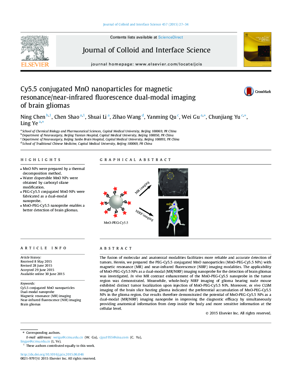
The fusion of molecular and anatomical modalities facilitates more reliable and accurate detection of tumors. Herein, we prepared the PEG-Cy5.5 conjugated MnO nanoparticles (MnO-PEG-Cy5.5 NPs) with magnetic resonance (MR) and near-infrared fluorescence (NIRF) imaging modalities. The applicability of MnO-PEG-Cy5.5 NPs as a dual-modal (MR/NIRF) imaging nanoprobe for the detection of brain gliomas was investigated. In vivo MR contrast enhancement of the MnO-PEG-Cy5.5 nanoprobe in the tumor region was demonstrated. Meanwhile, whole-body NIRF imaging of glioma bearing nude mouse exhibited distinct tumor localization upon injection of MnO-PEG-Cy5.5 NPs. Moreover, ex vivo CLSM imaging of the brain slice hosting glioma indicated the preferential accumulation of MnO-PEG-Cy5.5 NPs in the glioma region. Our results therefore demonstrated the potential of MnO-PEG-Cy5.5 NPs as a dual-modal (MR/NIRF) imaging nanoprobe in improving the diagnostic efficacy by simultaneously providing anatomical information from deep inside the body and more sensitive information at the cellular level.
Figure optionsDownload high-quality image (100 K)Download as PowerPoint slide
Journal: Journal of Colloid and Interface Science - Volume 457, 1 November 2015, Pages 27–34