| کد مقاله | کد نشریه | سال انتشار | مقاله انگلیسی | نسخه تمام متن |
|---|---|---|---|---|
| 865404 | 909665 | 2010 | 6 صفحه PDF | دانلود رایگان |
عنوان انگلیسی مقاله ISI
Phase Contrast Micro-Computed Tomography of Biological Sample at SSRF
دانلود مقاله + سفارش ترجمه
دانلود مقاله ISI انگلیسی
رایگان برای ایرانیان
موضوعات مرتبط
مهندسی و علوم پایه
سایر رشته های مهندسی
مهندسی (عمومی)
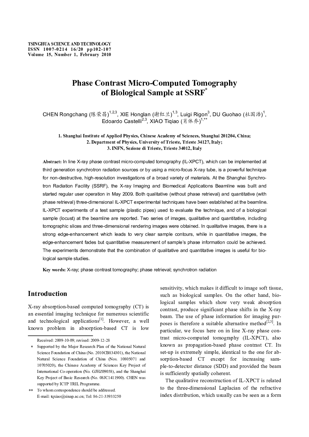
پیش نمایش صفحه اول مقاله

چکیده انگلیسی
In line X-ray phase contrast micro-computed tomography (IL-XPCT), which can be implemented at third generation synchrotron radiation sources or by using a micro-focus X-ray tube, is a powerful technique for non-destructive, high-resolution investigations of a broad variety of materials. At the Shanghai Synchrotron Radiation Facility (SSRF), the X-ray Imaging and Biomedical Applications Beamline was built and started regular user operation in May 2009. Both qualitative (without phase retrieval) and quantitative (with phase retrieval) three-dimensional IL-XPCT experimental techniques have been established at the beamline. IL-XPCT experiments of a test sample (plastic pipes) used to evaluate the technique, and of a biological sample (locust) at the beamline are reported. Two series of images, qualitative and quantitative, including tomographic slices and three-dimensional rendering images were obtained. In qualitative images, there is a strong edge-enhancement which leads to very clear sample contours, while in quantitative images, the edge-enhancement fades but quantitative measurement of sample's phase information could be achieved. The experiments demonstrate that the combination of qualitative and quantitative images is useful for biological sample studies.
ناشر
Database: Elsevier - ScienceDirect (ساینس دایرکت)
Journal: Tsinghua Science & Technology - Volume 15, Issue 1, February 2010, Pages 102-107
Journal: Tsinghua Science & Technology - Volume 15, Issue 1, February 2010, Pages 102-107
نویسندگان
Rongchang (éè£æ), Honglan (谢红å
°), Luigi Rigon, Guohao (æå½æµ©), Edoardo Castelli, Tiqiao (èä½ä¹),