| کد مقاله | کد نشریه | سال انتشار | مقاله انگلیسی | نسخه تمام متن |
|---|---|---|---|---|
| 9899186 | 1543766 | 2005 | 9 صفحه PDF | دانلود رایگان |
عنوان انگلیسی مقاله ISI
The detection of salmochelin and yersiniabactin in uropathogenic Escherichia coli strains by a novel hydrolysis-fluorescence-detection (HFD) method
دانلود مقاله + سفارش ترجمه
دانلود مقاله ISI انگلیسی
رایگان برای ایرانیان
موضوعات مرتبط
علوم زیستی و بیوفناوری
بیوشیمی، ژنتیک و زیست شناسی مولکولی
بیوشیمی، ژنتیک و زیست شناسی مولکولی (عمومی)
پیش نمایش صفحه اول مقاله

چکیده انگلیسی
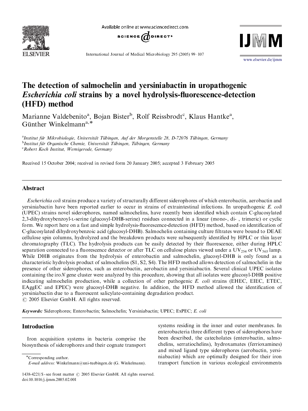
Escherichia coli strains produce a variety of structurally different siderophores of which enterobactin, aerobactin and yersiniabactin have been reported earlier to occur in strains of extraintestinal infections. In uropathogenic E. coli (UPEC) strains novel siderophores, named salmochelins, have recently been identified which contain C-glucosylated 2,3-dihydroxybenzoyl-l-serine (glucosyl-DHB-serine) residues connected in a linear (mono-, di- , trimeric) or cyclic form. We report here on a fast and simple hydrolysis-fluorescence-detection (HFD) method, based on identification of C-glucosylated dihydroxybenzoic acid (glucosyl-DHB). Salmochelin containing culture filtrates were bound to DEAE cellulose spin columns, hydrolyzed and the breakdown products were subsequently identified by HPLC or thin layer chromatography (TLC). The hydrolysis products can be easily detected by their fluorescence, either during HPLC separation connected to a fluorescence detector or after TLC on cellulose plates viewed under a UV254 or UV365 lamp. While DHB originates from the hydrolysis of enterobactin and salmochelin, glucosyl-DHB is only found as a characteristic hydrolysis product of salmochelins (S1, S2, S4). The HFD method allows detection of salmochelin in the presence of other siderophores, such as enterobactin, aerobactin and yersiniabactin. Several clinical UPEC isolates containing the iroN gene cluster were analyzed by this procedure, showing that all isolates were glucosyl-DHB positive indicating salmochelin production, while a collection of other pathogenic E. coli strains (EHEC, EIEC, ETEC, EAggEC and EPEC) were glucosyl-DHB negative. In addition, the HFD method allowed the identification of yersiniabactin due to a fluorescent salicylate-containing degradation product.
ناشر
Database: Elsevier - ScienceDirect (ساینس دایرکت)
Journal: International Journal of Medical Microbiology - Volume 295, Issue 2, 1 June 2005, Pages 99-107
Journal: International Journal of Medical Microbiology - Volume 295, Issue 2, 1 June 2005, Pages 99-107
نویسندگان
Marianne Valdebenito, Bojan Bister, Rolf Reissbrodt, Klaus Hantke, Günther Winkelmann,