| کد مقاله | کد نشریه | سال انتشار | مقاله انگلیسی | نسخه تمام متن |
|---|---|---|---|---|
| 10222558 | 1700997 | 2018 | 7 صفحه PDF | دانلود رایگان |
عنوان انگلیسی مقاله ISI
Isolated hepatic hemangiomatosis in 2 septuagenarians
دانلود مقاله + سفارش ترجمه
دانلود مقاله ISI انگلیسی
رایگان برای ایرانیان
موضوعات مرتبط
علوم پزشکی و سلامت
پزشکی و دندانپزشکی
رادیولوژی و تصویربرداری
پیش نمایش صفحه اول مقاله

چکیده انگلیسی
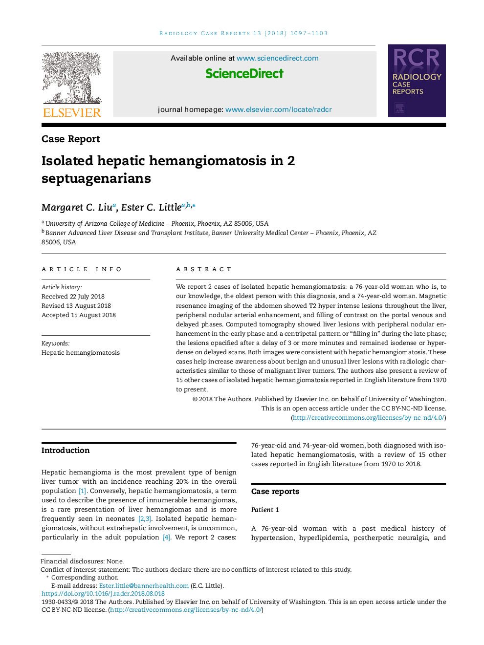
We report 2 cases of isolated hepatic hemangiomatosis: a 76-year-old woman who is, to our knowledge, the oldest person with this diagnosis, and a 74-year-old woman. Magnetic resonance imaging of the abdomen showed T2 hyper intense lesions throughout the liver, peripheral nodular arterial enhancement, and filling of contrast on the portal venous and delayed phases. Computed tomography showed liver lesions with peripheral nodular enhancement in the early phase and a centripetal pattern or “filling in” during the late phase; the lesions opacified after a delay of 3 or more minutes and remained isodense or hyperdense on delayed scans. Both images were consistent with hepatic hemangiomatosis. These cases help increase awareness about benign and unusual liver lesions with radiologic characteristics similar to those of malignant liver tumors. The authors also present a review of 15 other cases of isolated hepatic hemangiomatosis reported in English literature from 1970 to present.
ناشر
Database: Elsevier - ScienceDirect (ساینس دایرکت)
Journal: Radiology Case Reports - Volume 13, Issue 6, December 2018, Pages 1097-1103
Journal: Radiology Case Reports - Volume 13, Issue 6, December 2018, Pages 1097-1103
نویسندگان
Margaret C. Liu, Ester C. Little,