| کد مقاله | کد نشریه | سال انتشار | مقاله انگلیسی | نسخه تمام متن |
|---|---|---|---|---|
| 10228330 | 479 | 2013 | 11 صفحه PDF | دانلود رایگان |
عنوان انگلیسی مقاله ISI
In vivo fluorescence imaging for cancer diagnosis using receptor-targeted epidermal growth factor-based nanoprobe
دانلود مقاله + سفارش ترجمه
دانلود مقاله ISI انگلیسی
رایگان برای ایرانیان
کلمات کلیدی
موضوعات مرتبط
مهندسی و علوم پایه
مهندسی شیمی
بیو مهندسی (مهندسی زیستی)
پیش نمایش صفحه اول مقاله

چکیده انگلیسی
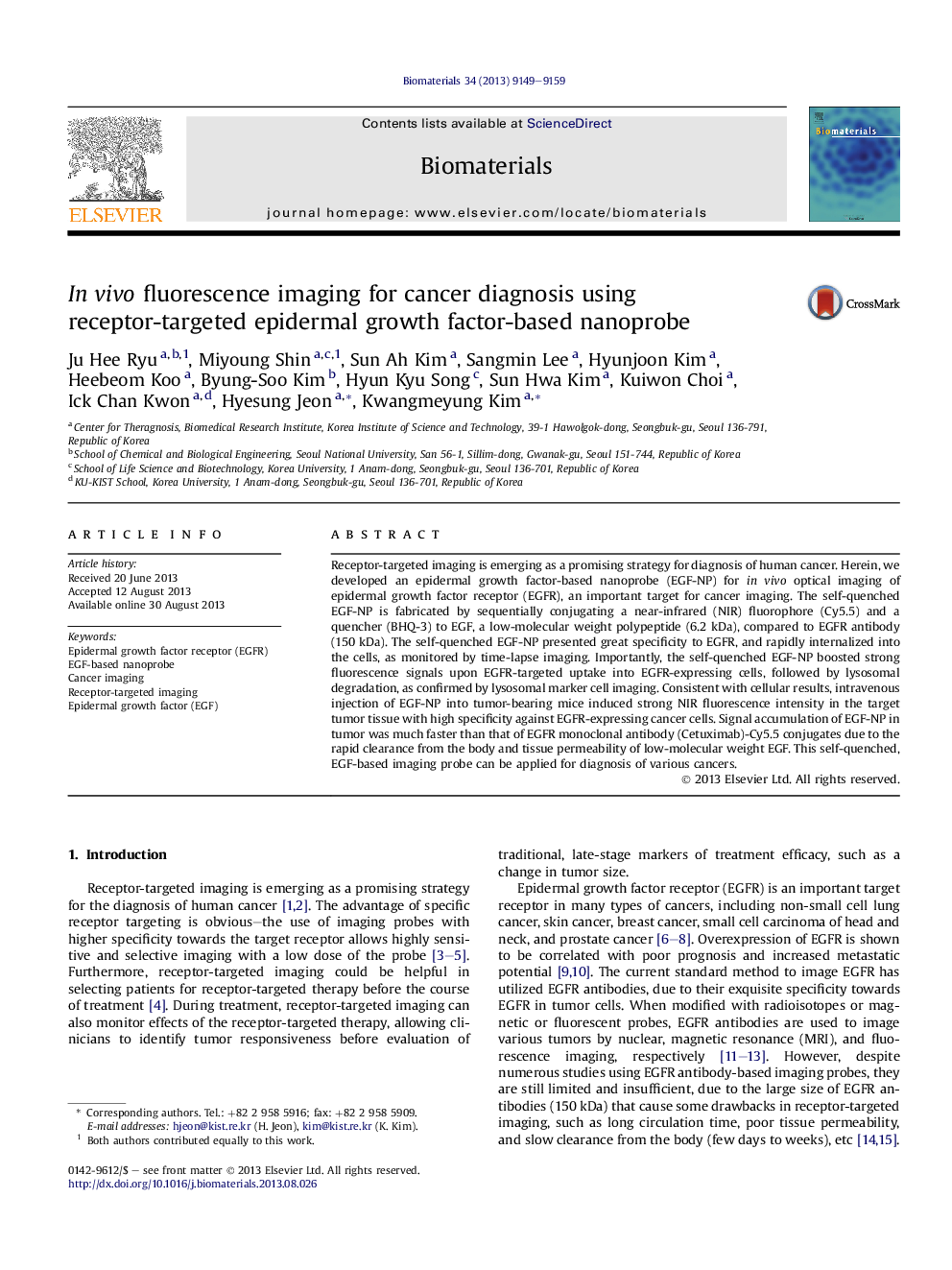
Receptor-targeted imaging is emerging as a promising strategy for diagnosis of human cancer. Herein, we developed an epidermal growth factor-based nanoprobe (EGF-NP) for in vivo optical imaging of epidermal growth factor receptor (EGFR), an important target for cancer imaging. The self-quenched EGF-NP is fabricated by sequentially conjugating a near-infrared (NIR) fluorophore (Cy5.5) and a quencher (BHQ-3) to EGF, a low-molecular weight polypeptide (6.2 kDa), compared to EGFR antibody (150 kDa). The self-quenched EGF-NP presented great specificity to EGFR, and rapidly internalized into the cells, as monitored by time-lapse imaging. Importantly, the self-quenched EGF-NP boosted strong fluorescence signals upon EGFR-targeted uptake into EGFR-expressing cells, followed by lysosomal degradation, as confirmed by lysosomal marker cell imaging. Consistent with cellular results, intravenous injection of EGF-NP into tumor-bearing mice induced strong NIR fluorescence intensity in the target tumor tissue with high specificity against EGFR-expressing cancer cells. Signal accumulation of EGF-NP in tumor was much faster than that of EGFR monoclonal antibody (Cetuximab)-Cy5.5 conjugates due to the rapid clearance from the body and tissue permeability of low-molecular weight EGF. This self-quenched, EGF-based imaging probe can be applied for diagnosis of various cancers.
ناشر
Database: Elsevier - ScienceDirect (ساینس دایرکت)
Journal: Biomaterials - Volume 34, Issue 36, December 2013, Pages 9149-9159
Journal: Biomaterials - Volume 34, Issue 36, December 2013, Pages 9149-9159
نویسندگان
Ju Hee Ryu, Miyoung Shin, Sun Ah Kim, Sangmin Lee, Hyunjoon Kim, Heebeom Koo, Byung-Soo Kim, Hyun Kyu Song, Sun Hwa Kim, Kuiwon Choi, Ick Chan Kwon, Hyesung Jeon, Kwangmeyung Kim,