| کد مقاله | کد نشریه | سال انتشار | مقاله انگلیسی | نسخه تمام متن |
|---|---|---|---|---|
| 2702045 | 1144511 | 2012 | 8 صفحه PDF | دانلود رایگان |
عنوان انگلیسی مقاله ISI
Computed Tomography Perfusion Imaging in Spectacular Shrinking Deficit
دانلود مقاله + سفارش ترجمه
دانلود مقاله ISI انگلیسی
رایگان برای ایرانیان
کلمات کلیدی
موضوعات مرتبط
علوم پزشکی و سلامت
پزشکی و دندانپزشکی
مغز و اعصاب بالینی
پیش نمایش صفحه اول مقاله

چکیده انگلیسی
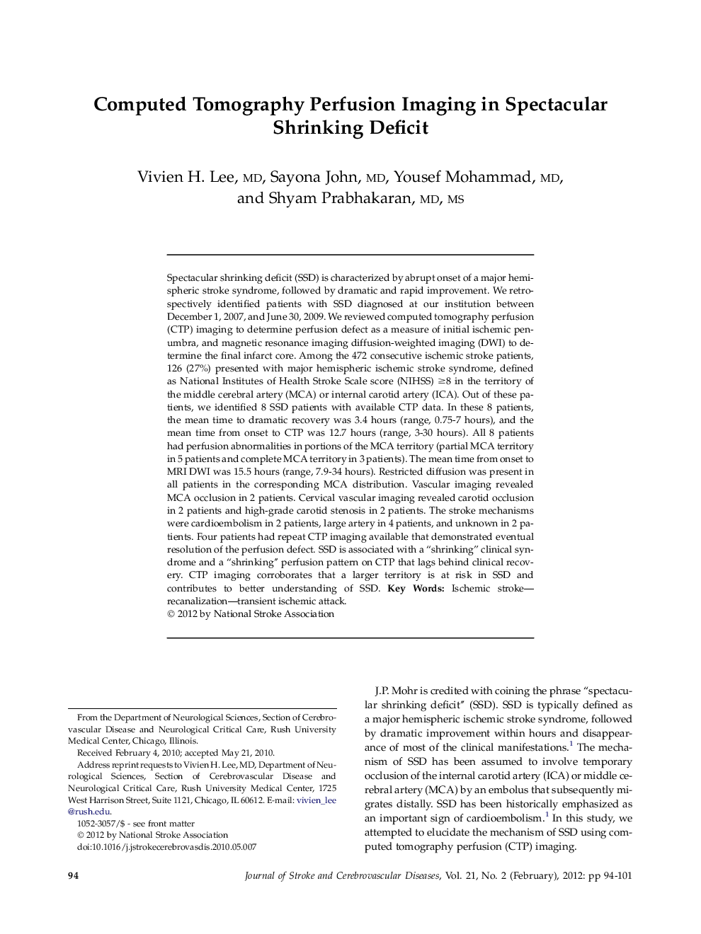
Spectacular shrinking deficit (SSD) is characterized by abrupt onset of a major hemispheric stroke syndrome, followed by dramatic and rapid improvement. We retrospectively identified patients with SSD diagnosed at our institution between December 1, 2007, and June 30, 2009. We reviewed computed tomography perfusion (CTP) imaging to determine perfusion defect as a measure of initial ischemic penumbra, and magnetic resonance imaging diffusion-weighted imaging (DWI) to determine the final infarct core. Among the 472 consecutive ischemic stroke patients, 126 (27%) presented with major hemispheric ischemic stroke syndrome, defined as National Institutes of Health Stroke Scale score (NIHSS) â¥8 in the territory of the middle cerebral artery (MCA) or internal carotid artery (ICA). Out of these patients, we identified 8 SSD patients with available CTP data. In these 8 patients, the mean time to dramatic recovery was 3.4 hours (range, 0.75-7 hours), and the mean time from onset to CTP was 12.7 hours (range, 3-30 hours). All 8 patients had perfusion abnormalities in portions of the MCA territory (partial MCA territory in 5 patients and complete MCA territory in 3 patients). The mean time from onset to MRI DWI was 15.5 hours (range, 7.9-34 hours). Restricted diffusion was present in all patients in the corresponding MCA distribution. Vascular imaging revealed MCA occlusion in 2 patients. Cervical vascular imaging revealed carotid occlusion in 2 patients and high-grade carotid stenosis in 2 patients. The stroke mechanisms were cardioembolism in 2 patients, large artery in 4 patients, and unknown in 2 patients. Four patients had repeat CTP imaging available that demonstrated eventual resolution of the perfusion defect. SSD is associated with a “shrinking” clinical syndrome and a “shrinking” perfusion pattern on CTP that lags behind clinical recovery. CTP imaging corroborates that a larger territory is at risk in SSD and contributes to better understanding of SSD.
ناشر
Database: Elsevier - ScienceDirect (ساینس دایرکت)
Journal: Journal of Stroke and Cerebrovascular Diseases - Volume 21, Issue 2, February 2012, Pages 94-101
Journal: Journal of Stroke and Cerebrovascular Diseases - Volume 21, Issue 2, February 2012, Pages 94-101
نویسندگان
Vivien H. MD, Sayona MD, Yousef MD, Shyam MD, MS,