| کد مقاله | کد نشریه | سال انتشار | مقاله انگلیسی | نسخه تمام متن |
|---|---|---|---|---|
| 3133712 | 1584203 | 2009 | 7 صفحه PDF | دانلود رایگان |

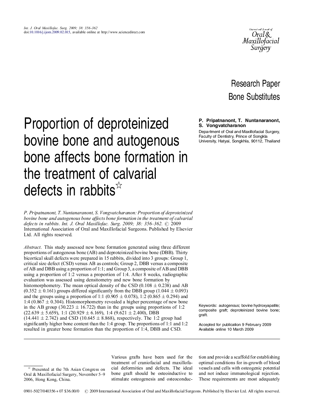
This study assessed new bone formation generated using three different proportions of autogenous bone (AB) and deproteinized bovine bone (DBB). Thirty bicortical skull defects were prepared in 15 rabbits, divided into 3 groups: Group 1, critical size defect (CSD) versus AB as controls; Group 2, DBB versus a composite of AB and DBB using a proportion of 1:1; and Group 3, a composite of AB and DBB using a proportion of 1:2 versus a proportion of 1:4. After 8 weeks, radiographic evaluation was assessed using densitometry and new bone formation by histomorphometry. The mean optical density of the CSD (0.108 ± 0.238) and AB (0.352 ± 0.161) groups differed significantly from the DBB group (1.044 ± 0.093) and the groups using a proportion of 1:1 (0.905 ± 0.078), 1:2 (0.865 ± 0.294) and 1:4 (0.867 ± 0.304). Histomorphometry revealed a higher percentage of new bone in the AB group (30.223 ± 16.722) than in the groups using proportions of 1:2 (22.639 ± 5.659), 1:1 (20.929 ± 6.169), 1:4 (9.621 ± 2.400), DBB (14.441 ± 2.742) and CSD (10.645 ± 8.868), respectively. The 1:2 group had significantly higher bone content than the 1:4 group. The proportions of 1:1 and 1:2 resulted in greater bone formation than the proportion of 1:4, DBB and CSD.
Journal: International Journal of Oral and Maxillofacial Surgery - Volume 38, Issue 4, April 2009, Pages 356–362