| کد مقاله | کد نشریه | سال انتشار | مقاله انگلیسی | نسخه تمام متن |
|---|---|---|---|---|
| 3133889 | 1584199 | 2009 | 4 صفحه PDF | دانلود رایگان |
عنوان انگلیسی مقاله ISI
Computed tomography morphology of the mandibular ramus at the lingual plane in patients with mandibular hyperplasia
دانلود مقاله + سفارش ترجمه
دانلود مقاله ISI انگلیسی
رایگان برای ایرانیان
کلمات کلیدی
موضوعات مرتبط
علوم پزشکی و سلامت
پزشکی و دندانپزشکی
دندانپزشکی، جراحی دهان و پزشکی
پیش نمایش صفحه اول مقاله

چکیده انگلیسی
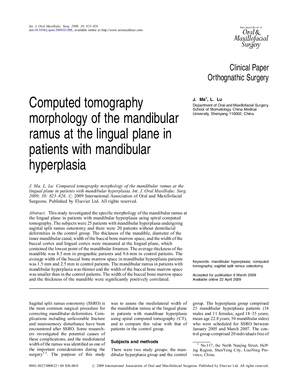
This study investigated the specific morphology of the mandibular ramus at the lingual plane in patients with mandibular hyperplasia using spiral computed tomography. The subjects were 25 patients with mandibular hyperplasia undergoing sagittal split ramus osteotomy and there were 20 patients without dentofacial deformities in the control group. The thickness of the mandible, diameter of the inner mandibular canal, width of the buccal bone marrow space, and the width of the buccal cortex and lingual cortex were measured at the lingual plane, which contained the lowest point of the mandibular foramen. The average thickness of the mandible was 8.5Â mm in prognathic patients and 9.6Â mm in control patients. The average width of the buccal bone marrow space in mandibular hyperplasia patients was 1.5Â mm and 2.5Â mm in control patients. The mandibular ramus in patients with mandibular hyperplasia was thinner and the width of the buccal bone marrow space was smaller than in the control patients. The width of the buccal bone marrow space and the thickness of the mandible were significantly positively correlated.
ناشر
Database: Elsevier - ScienceDirect (ساینس دایرکت)
Journal: International Journal of Oral and Maxillofacial Surgery - Volume 38, Issue 8, August 2009, Pages 823-826
Journal: International Journal of Oral and Maxillofacial Surgery - Volume 38, Issue 8, August 2009, Pages 823-826
نویسندگان
J. Ma, L. Lu,