| کد مقاله | کد نشریه | سال انتشار | مقاله انگلیسی | نسخه تمام متن |
|---|---|---|---|---|
| 3809645 | 1245561 | 2012 | 5 صفحه PDF | دانلود رایگان |
عنوان انگلیسی مقاله ISI
Interpretación de las pruebas de imagen en la patologÃa pancreática e interpretación clÃnica
دانلود مقاله + سفارش ترجمه
دانلود مقاله ISI انگلیسی
رایگان برای ایرانیان
کلمات کلیدی
Endoscopic retrograde cholangiopancreatography - cholangiopancreatography رتروگراد endoscopicPancreatic adenocarcinoma - آدنوکارسینوم پانکراسEcoendoscopia - اکوندیوسکوپیMagnetic resonance imaging - تصویربرداری رزونانس مغناطیسیcomputed tomography - توموگرافی کامپیوتری یا سی تی اسکن یا مقطعنگاری رایانهایResonancia magnética - رزونانس مغناطیسیPancreatitis - پانکراتیتColangiopancreatografía retrógrada endoscópica - کولونژیوپانکراپوگرافی رتروگراد endoscopic
موضوعات مرتبط
علوم پزشکی و سلامت
پزشکی و دندانپزشکی
پزشکی و دندانپزشکی (عمومی)
پیش نمایش صفحه اول مقاله

چکیده انگلیسی
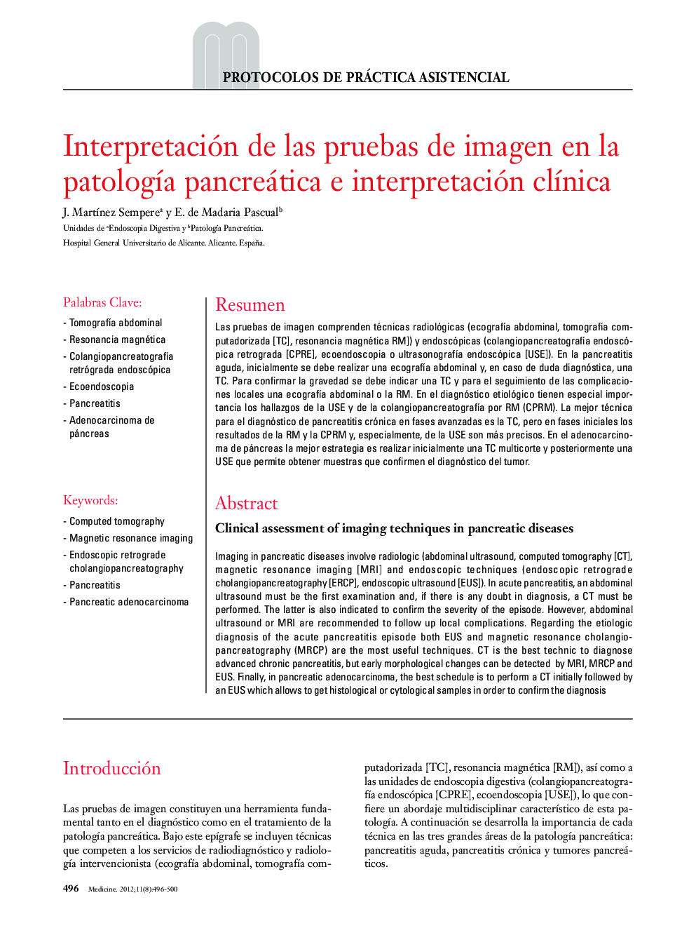
Imaging in pancreatic diseases involve radiologic (abdominal ultrasound, computed tomography [CT], magnetic resonance imaging [MRI] and endoscopic techniques (endoscopic retrograde cholangiopancreatography [ERCP], endoscopic ultrasound [EUS]). In acute pancreatitis, an abdominal ultrasound must be the first examination and, if there is any doubt in diagnosis, a CT must be performed. The latter is also indicated to confirm the severity of the episode. However, abdominal ultrasound or MRI are recommended to follow up local complications. Regarding the etiologic diagnosis of the acute pancreatitis episode both EUS and magnetic resonance cholangio-pancreatography (MRCP) are the most useful techniques. CT is the best technic to diagnose advanced chronic pancreatitis, but early morphological changes can be detected by MRI, MRCP and EUS. Finally, in pancreatic adenocarcinoma, the best schedule is to perform a CT initially followed by an EUS which allows to get histological or cytological samples in order to confirm the diagnosis
ناشر
Database: Elsevier - ScienceDirect (ساینس دایرکت)
Journal: Medicine - Programa de Formación Médica Continuada Acreditado - Volume 11, Issue 8, April 2012, Pages 496-500
Journal: Medicine - Programa de Formación Médica Continuada Acreditado - Volume 11, Issue 8, April 2012, Pages 496-500
نویسندگان
J. MartÃnez Sempere, E. de Madaria Pascual,