| کد مقاله | کد نشریه | سال انتشار | مقاله انگلیسی | نسخه تمام متن |
|---|---|---|---|---|
| 3812567 | 1597601 | 2006 | 5 صفحه PDF | دانلود رایگان |
عنوان انگلیسی مقاله ISI
Tumeur mélanocytaire primitive maligne radiculaire
دانلود مقاله + سفارش ترجمه
دانلود مقاله ISI انگلیسی
رایگان برای ایرانیان
موضوعات مرتبط
علوم زیستی و بیوفناوری
علم عصب شناسی
علوم اعصاب (عمومی)
پیش نمایش صفحه اول مقاله

چکیده انگلیسی
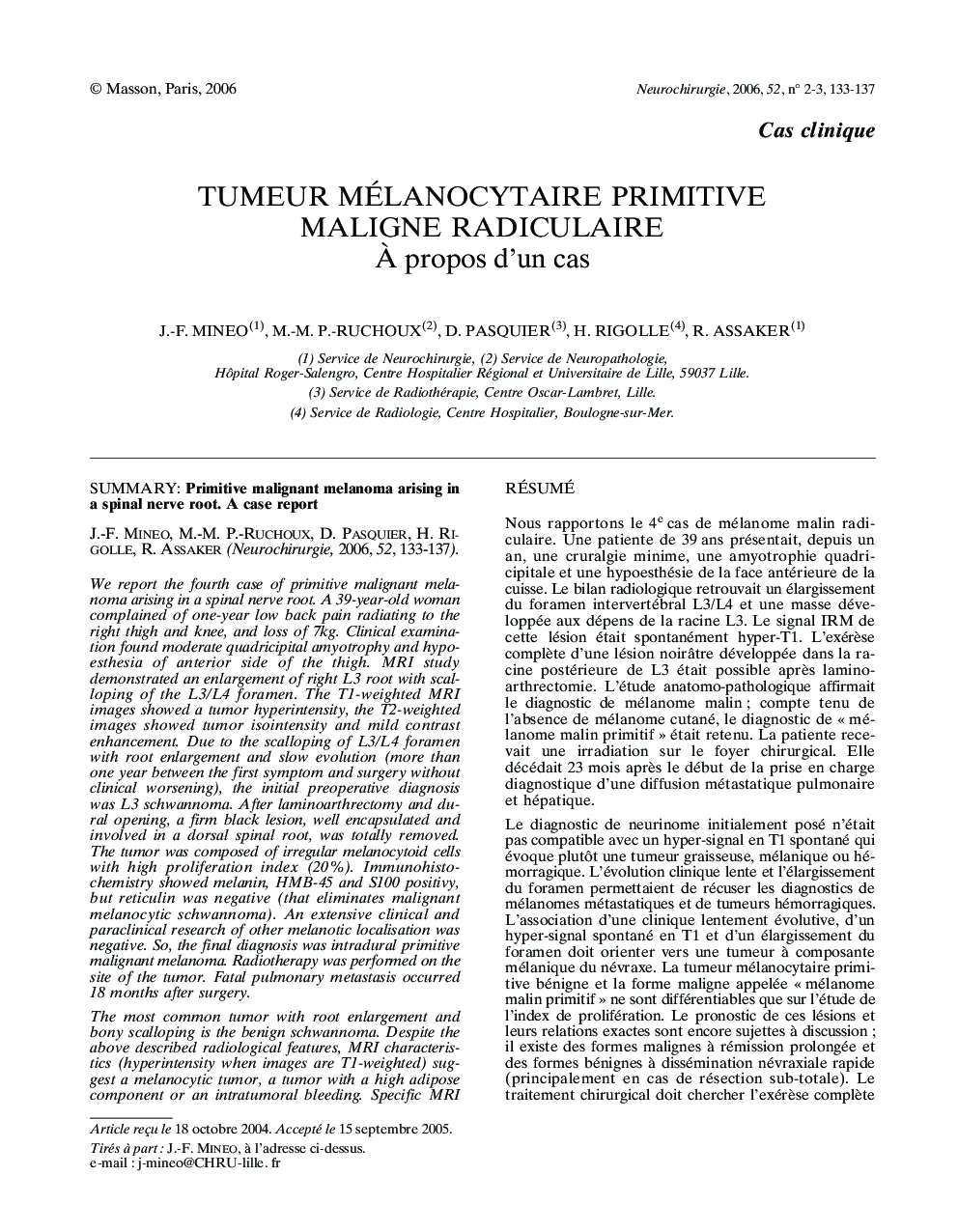
The most common tumor with root enlargement and bony scalloping is the benign schwannoma. Despite the above described radiological features, MRI characteristics (hyperintensity when images are T1-weighted) suggest a melanocytic tumor, a tumor with a high adipose component or an intratumoral bleeding. Specific MRI sequences can eliminate adipose tissue tumor, but diagnosis between melanin and methemoglobin is still difficult. According to the index of proliferation, a primitive central melanocytic lesion can be a meningeal melanocytoma (considered as benign) or a primitive malignant melanoma. These tumors show identical protein expressions in immunohistochemistry, and their prognosis is very variable (some long-term remissions are reported for malignant melanomas and fast disseminations are described for meningeal melanocytomas treated by sub-total surgery). The L3/L4 foramen scalloping is unusual for a malignant lesion with theoretic high-speed development. The other 3 patients (reported in the literature) survive more than 3 years. The histological features of malignant lesion with benign clinical features lead to interrogation upon the actual pathologic classification.
ناشر
Database: Elsevier - ScienceDirect (ساینس دایرکت)
Journal: Neurochirurgie - Volume 52, Issues 2â3, Part 1, June 2006, Pages 133-137
Journal: Neurochirurgie - Volume 52, Issues 2â3, Part 1, June 2006, Pages 133-137
نویسندگان
J.-F. Mineo, M.-M. P.-Ruchoux, D. Pasquier, H. Rigolle, R. Assaker,