| کد مقاله | کد نشریه | سال انتشار | مقاله انگلیسی | نسخه تمام متن |
|---|---|---|---|---|
| 4221609 | 1281626 | 2012 | 4 صفحه PDF | دانلود رایگان |
عنوان انگلیسی مقاله ISI
Ultrasound and MR images of prenatally diagnosed bilateral congenital diaphragmatic hernia, a rare variation of CDH
دانلود مقاله + سفارش ترجمه
دانلود مقاله ISI انگلیسی
رایگان برای ایرانیان
کلمات کلیدی
موضوعات مرتبط
علوم پزشکی و سلامت
پزشکی و دندانپزشکی
رادیولوژی و تصویربرداری
پیش نمایش صفحه اول مقاله

چکیده انگلیسی
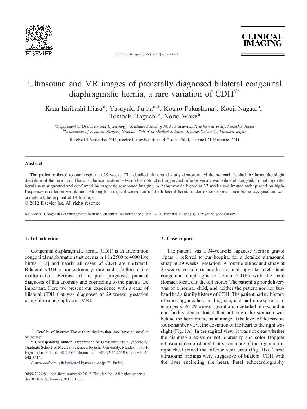
The patient referred to our hospital at 29 weeks. The detailed ultrasound study demonstrated the stomach behind the heart, the slight deviation of the heart, and the vascular connection between the right chest organ and inferior vena cava. Bilateral congenital diaphragmatic hernia was suggested and confirmed by magnetic resonance imaging. A baby was delivered at 37 weeks and immediately placed on high-frequency oscillation ventilation. Although a surgical correction of the bilateral hernia under extracorporeal membrane oxygenation was completed, he expired at 14 h of age.
ناشر
Database: Elsevier - ScienceDirect (ساینس دایرکت)
Journal: Clinical Imaging - Volume 36, Issue 5, September–October 2012, Pages 639–642
Journal: Clinical Imaging - Volume 36, Issue 5, September–October 2012, Pages 639–642
نویسندگان
Kana Ishibashi Hiasa, Yasuyuki Fujita, Kotaro Fukushima, Kouji Nagata, Tomoaki Taguchi, Norio Wake,