| کد مقاله | کد نشریه | سال انتشار | مقاله انگلیسی | نسخه تمام متن |
|---|---|---|---|---|
| 4223261 | 1281686 | 2006 | 5 صفحه PDF | دانلود رایگان |
عنوان انگلیسی مقاله ISI
Imaging and pathological correlation of soft-tissue chondroma: A serial five-case study and literature review
دانلود مقاله + سفارش ترجمه
دانلود مقاله ISI انگلیسی
رایگان برای ایرانیان
موضوعات مرتبط
علوم پزشکی و سلامت
پزشکی و دندانپزشکی
رادیولوژی و تصویربرداری
پیش نمایش صفحه اول مقاله

چکیده انگلیسی
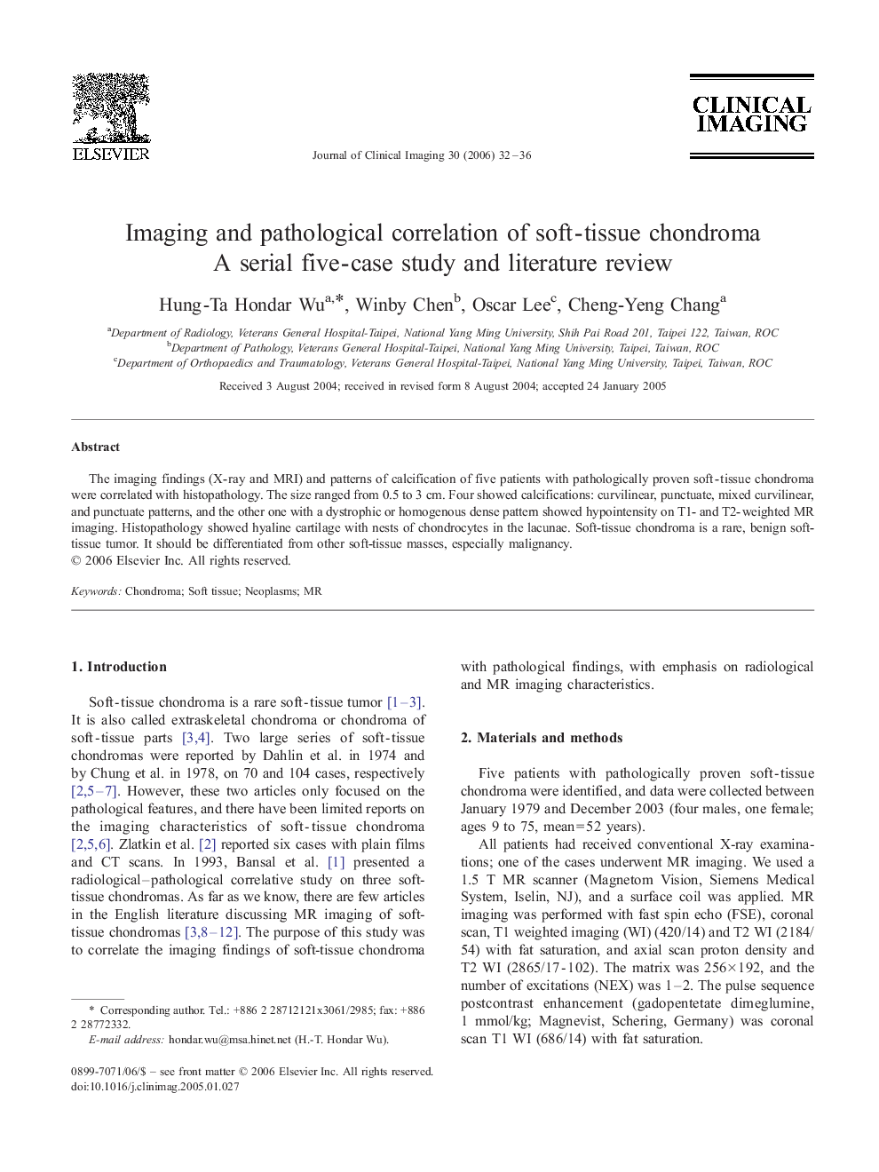
The imaging findings (X-ray and MRI) and patterns of calcification of five patients with pathologically proven soft-tissue chondroma were correlated with histopathology. The size ranged from 0.5 to 3 cm. Four showed calcifications: curvilinear, punctuate, mixed curvilinear, and punctuate patterns, and the other one with a dystrophic or homogenous dense pattern showed hypointensity on T1- and T2-weighted MR imaging. Histopathology showed hyaline cartilage with nests of chondrocytes in the lacunae. Soft-tissue chondroma is a rare, benign soft-tissue tumor. It should be differentiated from other soft-tissue masses, especially malignancy.
ناشر
Database: Elsevier - ScienceDirect (ساینس دایرکت)
Journal: Clinical Imaging - Volume 30, Issue 1, January–February 2006, Pages 32–36
Journal: Clinical Imaging - Volume 30, Issue 1, January–February 2006, Pages 32–36
نویسندگان
Hung-Ta Hondar Wu, Winby Chen, Oscar Lee, Cheng-Yeng Chang,