| کد مقاله | کد نشریه | سال انتشار | مقاله انگلیسی | نسخه تمام متن |
|---|---|---|---|---|
| 4248492 | 1283742 | 2007 | 9 صفحه PDF | دانلود رایگان |
عنوان انگلیسی مقاله ISI
Primary Synovial Sarcoma of the Lung
دانلود مقاله + سفارش ترجمه
دانلود مقاله ISI انگلیسی
رایگان برای ایرانیان
موضوعات مرتبط
علوم پزشکی و سلامت
پزشکی و دندانپزشکی
رادیولوژی و تصویربرداری
پیش نمایش صفحه اول مقاله

چکیده انگلیسی
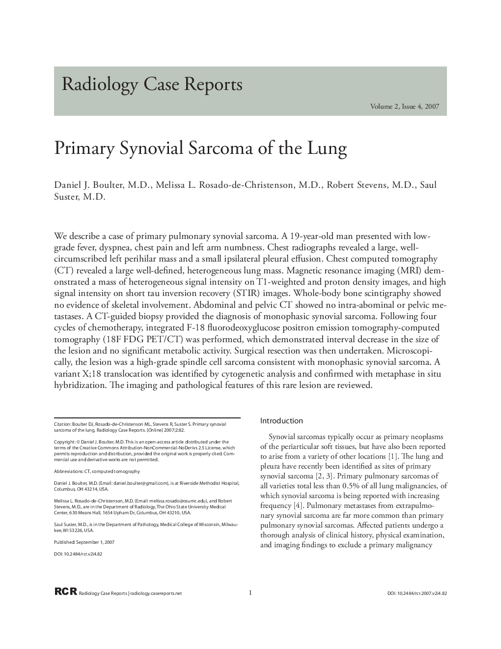
We describe a case of primary pulmonary synovial sarcoma. A 19-year-old man presented with low-grade fever, dyspnea, chest pain and left arm numbness. Chest radiographs revealed a large, well-circumscribed left perihilar mass and a small ipsilateral pleural effusion. Chest computed tomography (CT) revealed a large well-defined, heterogeneous lung mass. Magnetic resonance imaging (MRI) demonstrated a mass of heterogeneous signal intensity on T1-weighted and proton density images, and high signal intensity on short tau inversion recovery (STIR) images. Whole-body bone scintigraphy showed no evidence of skeletal involvement. Abdominal and pelvic CT showed no intra-abominal or pelvic metastases. A CT-guided biopsy provided the diagnosis of monophasic synovial sarcoma. Following four cycles of chemotherapy, integrated F-18 fluorodeoxyglucose positron emission tomography-computed tomography (18F FDG PET/CT) was performed, which demonstrated interval decrease in the size of the lesion and no significant metabolic activity. Surgical resection was then undertaken. Microscopically, the lesion was a high-grade spindle cell sarcoma consistent with monophasic synovial sarcoma. A variant X;18 translocation was identified by cytogenetic analysis and confirmed with metaphase in situ hybridization. The imaging and pathological features of this rare lesion are reviewed.
ناشر
Database: Elsevier - ScienceDirect (ساینس دایرکت)
Journal: Radiology Case Reports - Volume 2, Issue 4, 2007, 82
Journal: Radiology Case Reports - Volume 2, Issue 4, 2007, 82
نویسندگان
Daniel J. M.D., Melissa L. M.D., Robert M.D., Saul M.D.,