| کد مقاله | کد نشریه | سال انتشار | مقاله انگلیسی | نسخه تمام متن |
|---|---|---|---|---|
| 4248548 | 1283746 | 2007 | 4 صفحه PDF | دانلود رایگان |
عنوان انگلیسی مقاله ISI
Hemangioma of the Ilium Simulating an Aggressive Bone Lesion on Imaging
دانلود مقاله + سفارش ترجمه
دانلود مقاله ISI انگلیسی
رایگان برای ایرانیان
کلمات کلیدی
موضوعات مرتبط
علوم پزشکی و سلامت
پزشکی و دندانپزشکی
رادیولوژی و تصویربرداری
پیش نمایش صفحه اول مقاله

چکیده انگلیسی
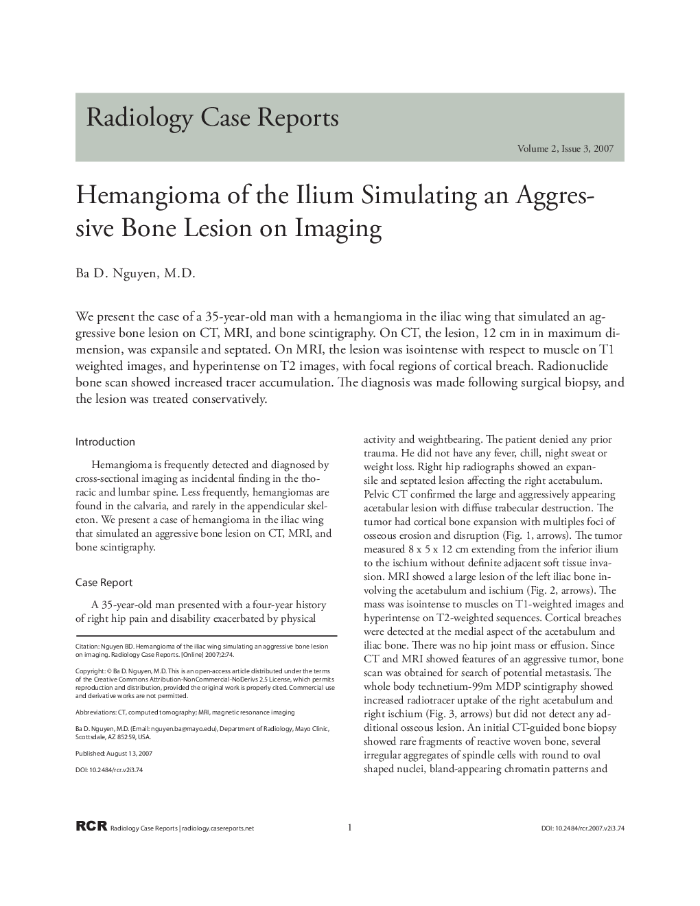
We present the case of a 35-year-old man with a hemangioma in the iliac wing that simulated an aggressive bone lesion on CT, MRI, and bone scintigraphy. On CT, the lesion, 12 cm in in maximum dimension, was expansile and septated. On MRI, the lesion was isointense with respect to muscle on T1 weighted images, and hyperintense on T2 images, with focal regions of cortical breach. Radionuclide bone scan showed increased tracer accumulation. The diagnosis was made following surgical biopsy, and the lesion was treated conservatively.
ناشر
Database: Elsevier - ScienceDirect (ساینس دایرکت)
Journal: Radiology Case Reports - Volume 2, Issue 3, 2007, 74
Journal: Radiology Case Reports - Volume 2, Issue 3, 2007, 74
نویسندگان
Ba D. M.D.,