| کد مقاله | کد نشریه | سال انتشار | مقاله انگلیسی | نسخه تمام متن |
|---|---|---|---|---|
| 4327359 | 1614120 | 2010 | 11 صفحه PDF | دانلود رایگان |
عنوان انگلیسی مقاله ISI
Microglial response to murine leukemia virus-induced encephalopathy is a good indicator of neuronal perturbations
دانلود مقاله + سفارش ترجمه
دانلود مقاله ISI انگلیسی
رایگان برای ایرانیان
کلمات کلیدی
موضوعات مرتبط
علوم زیستی و بیوفناوری
علم عصب شناسی
علوم اعصاب (عمومی)
پیش نمایش صفحه اول مقاله

چکیده انگلیسی
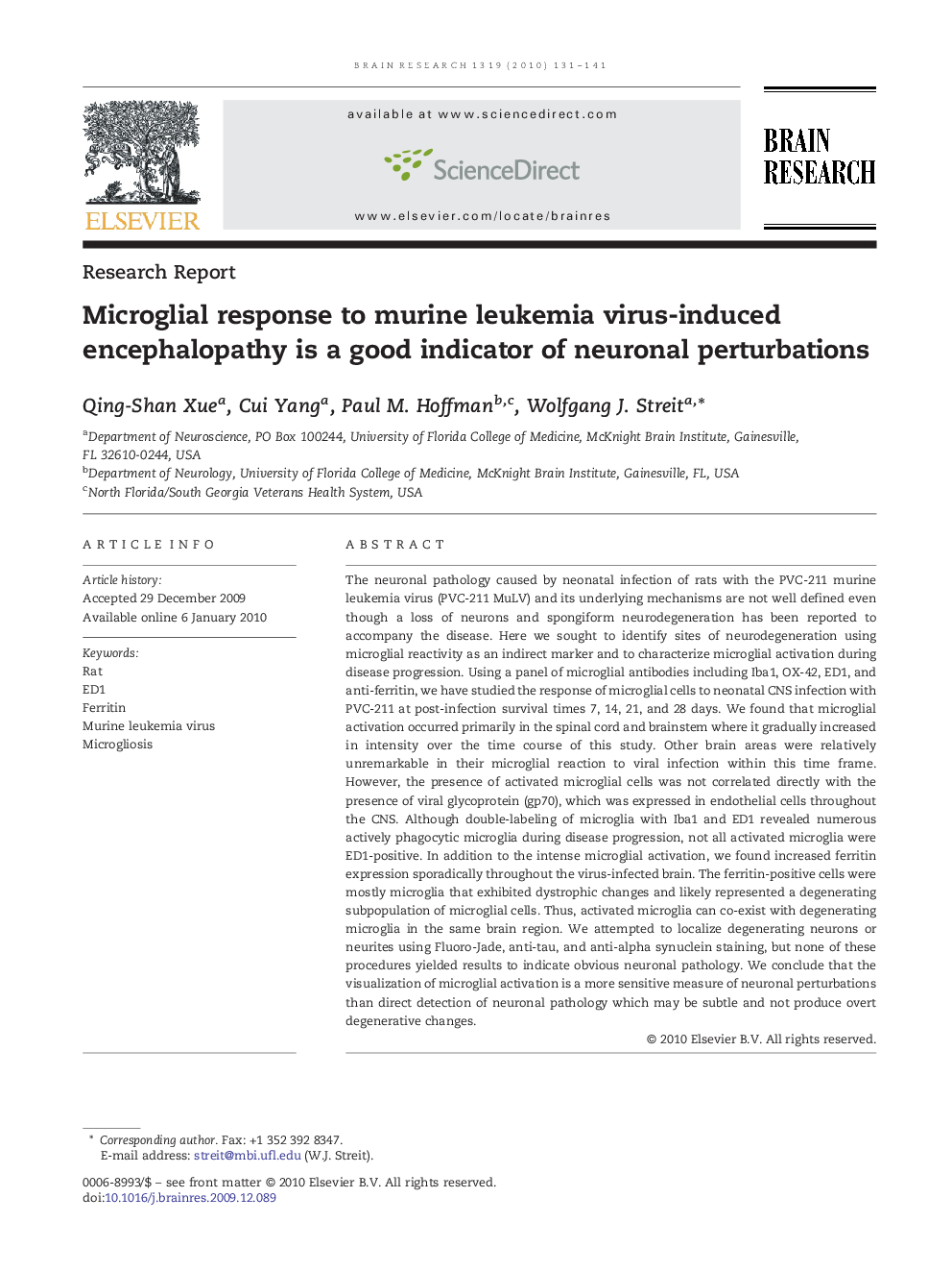
The neuronal pathology caused by neonatal infection of rats with the PVC-211 murine leukemia virus (PVC-211 MuLV) and its underlying mechanisms are not well defined even though a loss of neurons and spongiform neurodegeneration has been reported to accompany the disease. Here we sought to identify sites of neurodegeneration using microglial reactivity as an indirect marker and to characterize microglial activation during disease progression. Using a panel of microglial antibodies including Iba1, OX-42, ED1, and anti-ferritin, we have studied the response of microglial cells to neonatal CNS infection with PVC-211 at post-infection survival times 7, 14, 21, and 28 days. We found that microglial activation occurred primarily in the spinal cord and brainstem where it gradually increased in intensity over the time course of this study. Other brain areas were relatively unremarkable in their microglial reaction to viral infection within this time frame. However, the presence of activated microglial cells was not correlated directly with the presence of viral glycoprotein (gp70), which was expressed in endothelial cells throughout the CNS. Although double-labeling of microglia with Iba1 and ED1 revealed numerous actively phagocytic microglia during disease progression, not all activated microglia were ED1-positive. In addition to the intense microglial activation, we found increased ferritin expression sporadically throughout the virus-infected brain. The ferritin-positive cells were mostly microglia that exhibited dystrophic changes and likely represented a degenerating subpopulation of microglial cells. Thus, activated microglia can co-exist with degenerating microglia in the same brain region. We attempted to localize degenerating neurons or neurites using Fluoro-Jade, anti-tau, and anti-alpha synuclein staining, but none of these procedures yielded results to indicate obvious neuronal pathology. We conclude that the visualization of microglial activation is a more sensitive measure of neuronal perturbations than direct detection of neuronal pathology which may be subtle and not produce overt degenerative changes.
ناشر
Database: Elsevier - ScienceDirect (ساینس دایرکت)
Journal: Brain Research - Volume 1319, 10 March 2010, Pages 131-141
Journal: Brain Research - Volume 1319, 10 March 2010, Pages 131-141
نویسندگان
Qing-Shan Xue, Cui Yang, Paul M. Hoffman, Wolfgang J. Streit,