| کد مقاله | کد نشریه | سال انتشار | مقاله انگلیسی | نسخه تمام متن |
|---|---|---|---|---|
| 5579589 | 1404121 | 2017 | 18 صفحه PDF | دانلود رایگان |
عنوان انگلیسی مقاله ISI
Magnetic Resonance Imaging of Pelvic Masses: A Compartmental Approach
ترجمه فارسی عنوان
تصویربرداری رزونانس مغناطیسی توده های نخاعی: رویکرد مجتمع
دانلود مقاله + سفارش ترجمه
دانلود مقاله ISI انگلیسی
رایگان برای ایرانیان
موضوعات مرتبط
علوم پزشکی و سلامت
پزشکی و دندانپزشکی
رادیولوژی و تصویربرداری
چکیده انگلیسی
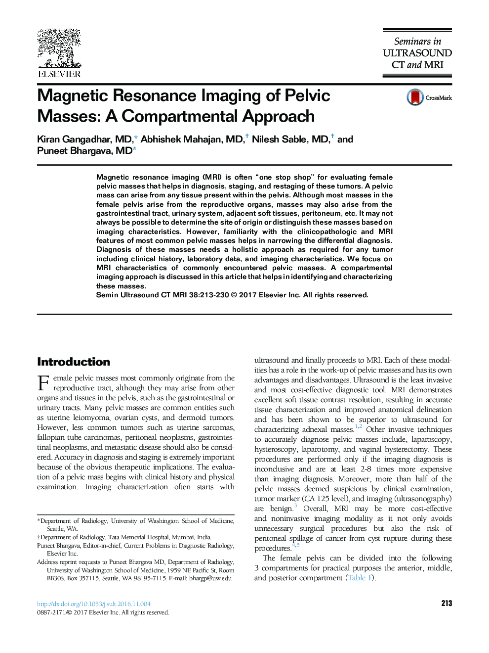
Magnetic resonance imaging (MRI) is often “one stop shop” for evaluating female pelvic masses that helps in diagnosis, staging, and restaging of these tumors. A pelvic mass can arise from any tissue present within the pelvis. Although most masses in the female pelvis arise from the reproductive organs, masses may also arise from the gastrointestinal tract, urinary system, adjacent soft tissues, peritoneum, etc. It may not always be possible to determine the site of origin or distinguish these masses based on imaging characteristics. However, familiarity with the clinicopathologic and MRI features of most common pelvic masses helps in narrowing the differential diagnosis. Diagnosis of these masses needs a holistic approach as required for any tumor including clinical history, laboratory data, and imaging characteristics. We focus on MRI characteristics of commonly encountered pelvic masses. A compartmental imaging approach is discussed in this article that helps in identifying and characterizing these masses.
ناشر
Database: Elsevier - ScienceDirect (ساینس دایرکت)
Journal: Seminars in Ultrasound, CT and MRI - Volume 38, Issue 3, June 2017, Pages 213-230
Journal: Seminars in Ultrasound, CT and MRI - Volume 38, Issue 3, June 2017, Pages 213-230
نویسندگان
Kiran MD, Abhishek MD, Nilesh MD, Puneet MD,
