| کد مقاله | کد نشریه | سال انتشار | مقاله انگلیسی | نسخه تمام متن |
|---|---|---|---|---|
| 5714235 | 1605712 | 2017 | 4 صفحه PDF | دانلود رایگان |
عنوان انگلیسی مقاله ISI
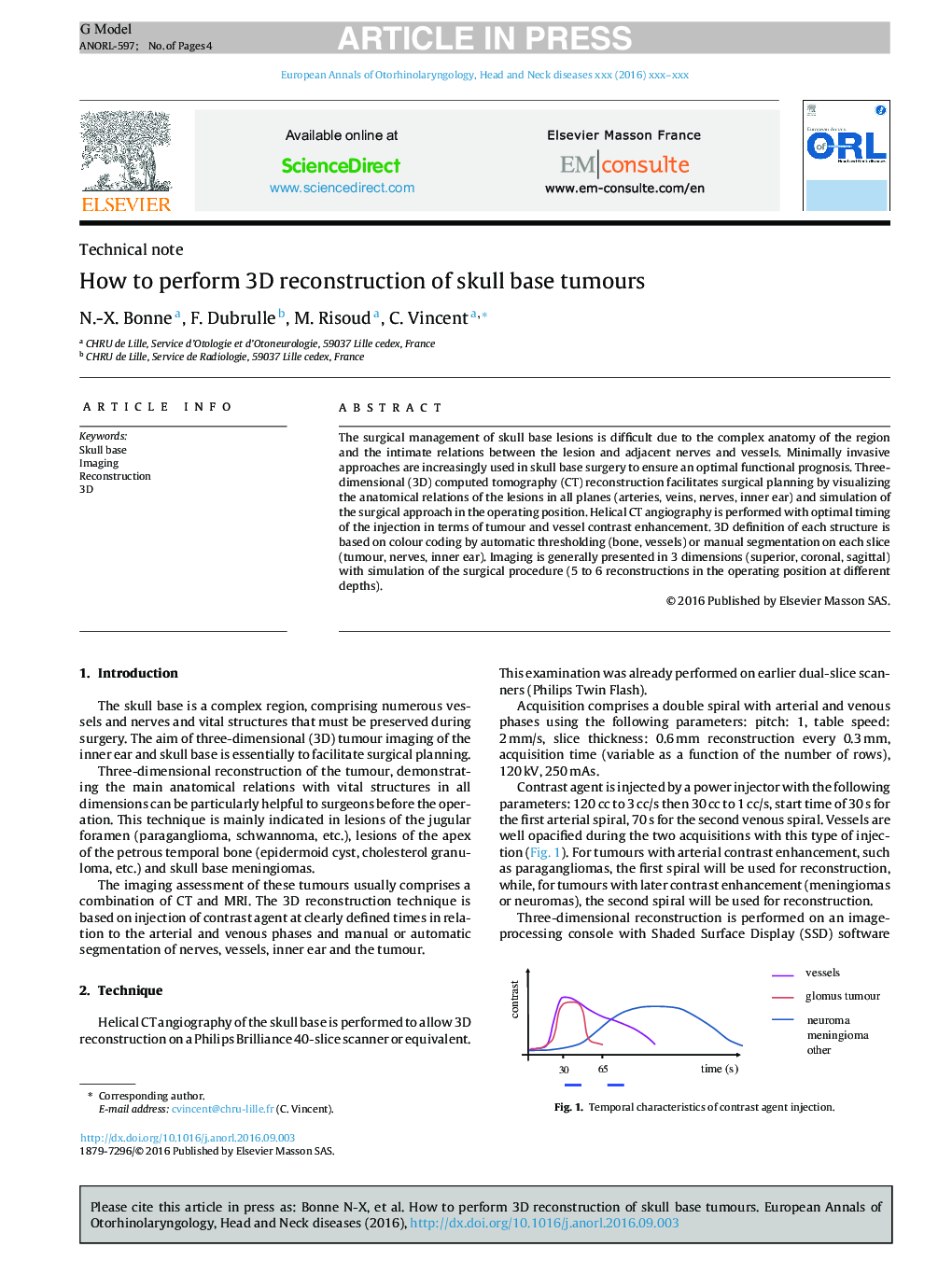
How to perform 3D reconstruction of skull base tumours
ترجمه فارسی عنوان
نحوه انجام بازسازی سه بعدی تومورهای پایه جمجمه
دانلود مقاله + سفارش ترجمه
دانلود مقاله ISI انگلیسی
رایگان برای ایرانیان
موضوعات مرتبط
علوم پزشکی و سلامت
پزشکی و دندانپزشکی
بیماری های گوش و جراحی پلاستیک صورت
چکیده انگلیسی
The surgical management of skull base lesions is difficult due to the complex anatomy of the region and the intimate relations between the lesion and adjacent nerves and vessels. Minimally invasive approaches are increasingly used in skull base surgery to ensure an optimal functional prognosis. Three-dimensional (3D) computed tomography (CT) reconstruction facilitates surgical planning by visualizing the anatomical relations of the lesions in all planes (arteries, veins, nerves, inner ear) and simulation of the surgical approach in the operating position. Helical CT angiography is performed with optimal timing of the injection in terms of tumour and vessel contrast enhancement. 3D definition of each structure is based on colour coding by automatic thresholding (bone, vessels) or manual segmentation on each slice (tumour, nerves, inner ear). Imaging is generally presented in 3 dimensions (superior, coronal, sagittal) with simulation of the surgical procedure (5 to 6 reconstructions in the operating position at different depths).
ناشر
Database: Elsevier - ScienceDirect (ساینس دایرکت)
Journal: European Annals of Otorhinolaryngology, Head and Neck Diseases - Volume 134, Issue 2, April 2017, Pages 117-120
Journal: European Annals of Otorhinolaryngology, Head and Neck Diseases - Volume 134, Issue 2, April 2017, Pages 117-120
نویسندگان
N.-X. Bonne, F. Dubrulle, M. Risoud, C. Vincent,
