| کد مقاله | کد نشریه | سال انتشار | مقاله انگلیسی | نسخه تمام متن |
|---|---|---|---|---|
| 6059284 | 1199177 | 2012 | 9 صفحه PDF | دانلود رایگان |
عنوان انگلیسی مقاله ISI
The clinical and radiographic characteristics of condylar osteochondroma
دانلود مقاله + سفارش ترجمه
دانلود مقاله ISI انگلیسی
رایگان برای ایرانیان
موضوعات مرتبط
علوم پزشکی و سلامت
پزشکی و دندانپزشکی
دندانپزشکی، جراحی دهان و پزشکی
پیش نمایش صفحه اول مقاله

چکیده انگلیسی
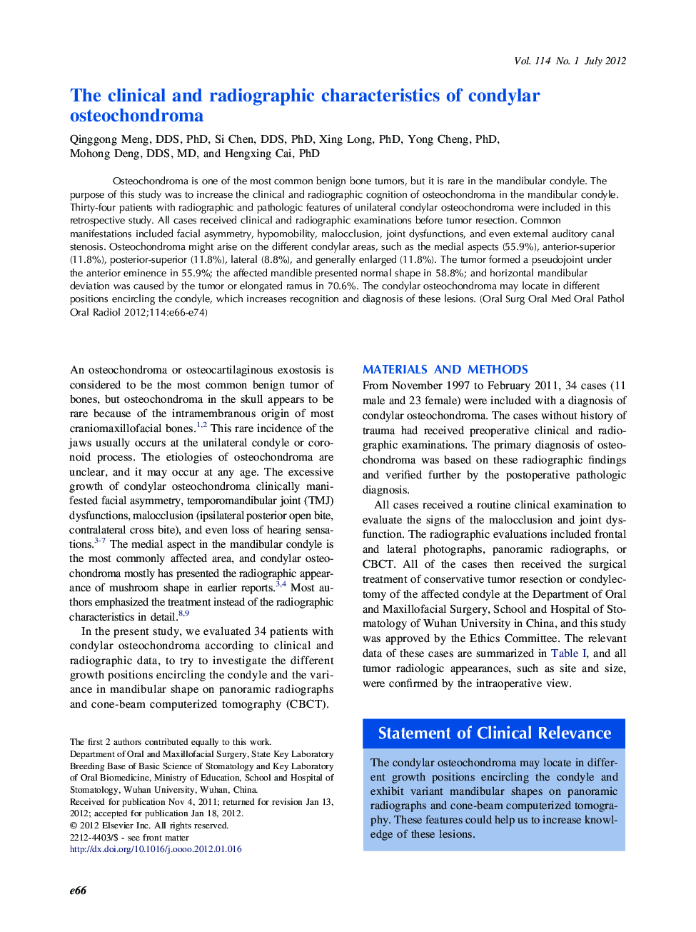
Osteochondroma is one of the most common benign bone tumors, but it is rare in the mandibular condyle. The purpose of this study was to increase the clinical and radiographic cognition of osteochondroma in the mandibular condyle. Thirty-four patients with radiographic and pathologic features of unilateral condylar osteochondroma were included in this retrospective study. All cases received clinical and radiographic examinations before tumor resection. Common manifestations included facial asymmetry, hypomobility, malocclusion, joint dysfunctions, and even external auditory canal stenosis. Osteochondroma might arise on the different condylar areas, such as the medial aspects (55.9%), anterior-superior (11.8%), posterior-superior (11.8%), lateral (8.8%), and generally enlarged (11.8%). The tumor formed a pseudojoint under the anterior eminence in 55.9%; the affected mandible presented normal shape in 58.8%; and horizontal mandibular deviation was caused by the tumor or elongated ramus in 70.6%. The condylar osteochondroma may locate in different positions encircling the condyle, which increases recognition and diagnosis of these lesions.
ناشر
Database: Elsevier - ScienceDirect (ساینس دایرکت)
Journal: Oral Surgery, Oral Medicine, Oral Pathology and Oral Radiology - Volume 114, Issue 1, July 2012, Pages e66-e74
Journal: Oral Surgery, Oral Medicine, Oral Pathology and Oral Radiology - Volume 114, Issue 1, July 2012, Pages e66-e74
نویسندگان
Qinggong DDS, PhD, Si DDS, PhD, Xing PhD, Yong PhD, Mohong DDS, MD, Hengxing PhD,