| کد مقاله | کد نشریه | سال انتشار | مقاله انگلیسی | نسخه تمام متن |
|---|---|---|---|---|
| 6268080 | 1614612 | 2016 | 11 صفحه PDF | دانلود رایگان |
- User-friendly semi-automatic perivascular spaces segmentation method is efficient in quantifying PVS from conventional T2-weighted MRI.
- Semi-automatically determined perivascular spaces count and volume agree with visual ratings.
- Semi-automatic perivascular spaces assessment proves useful for clinical studies and MRI protocols.
- Rigorous statistical analysis evaluating the new method on a longitudinal stroke sample.
- Potential for use in studies of cerebral small vessel disease and in a range of other neurological diseases.
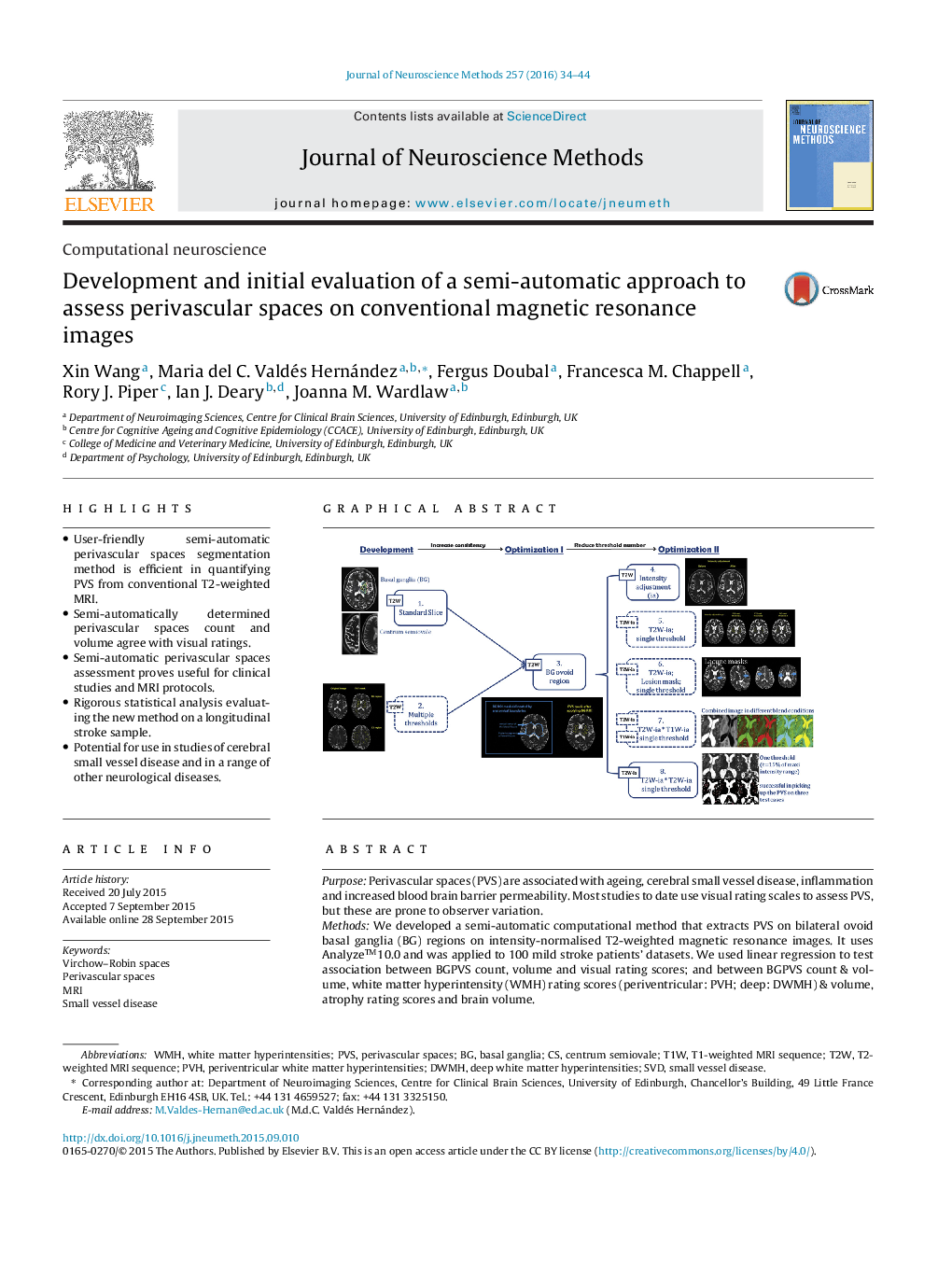
PurposePerivascular spaces (PVS) are associated with ageing, cerebral small vessel disease, inflammation and increased blood brain barrier permeability. Most studies to date use visual rating scales to assess PVS, but these are prone to observer variation.MethodsWe developed a semi-automatic computational method that extracts PVS on bilateral ovoid basal ganglia (BG) regions on intensity-normalised T2-weighted magnetic resonance images. It uses Analyzeâ¢10.0 and was applied to 100 mild stroke patients' datasets. We used linear regression to test association between BGPVS count, volume and visual rating scores; and between BGPVS count & volume, white matter hyperintensity (WMH) rating scores (periventricular: PVH; deep: DWMH) & volume, atrophy rating scores and brain volume.ResultsIn the 100 patients WMH ranged from 0.4 to 119 ml, and total brain tissue volume from 0.65 to 1.45 l. BGPVS volume increased with BGPVS count (67.27, 95%CI [57.93 to 76.60], p < 0.001). BGPVS count was positively associated with WMH visual rating (PVH: 2.20, 95%CI [1.22 to 3.18], p < 0.001; DWMH: 1.92, 95%CI [0.99 to 2.85], p < 0.001), WMH volume (0.065, 95%CI [0.034 to 0.096], p < 0.001), and whole brain atrophy visual rating (1.01, 95%CI [0.49 to 1.53], p < 0.001). BGPVS count increased as brain volume (as % of ICV) decreased (â0.33, 95%CI [â0.53 to â0.13], p = 0.002).Comparison with existing methodBGPVS count and volume increased with the overall increase of BGPVS visual scores (2.11, 95%CI [1.36 to 2.86] for count and 0.022, 95%CI [0.012 to 0.031] for volume, p < 0.001). Distributions for PVS count and visual scores were also similar.ConclusionsThis semi-automatic method is applicable to clinical protocols and offers quantitative surrogates for PVS load. It shows good agreement with a visual rating scale and confirmed that BGPVS are associated with WMH and atrophy measurements.
Journal: Journal of Neuroscience Methods - Volume 257, 15 January 2016, Pages 34-44
