| کد مقاله | کد نشریه | سال انتشار | مقاله انگلیسی | نسخه تمام متن |
|---|---|---|---|---|
| 7239229 | 1471152 | 2017 | 7 صفحه PDF | دانلود رایگان |
عنوان انگلیسی مقاله ISI
Treatment of multifocal central nervous system AIDS-related Epstein Barr virus-associated malignant myopericytoma with bevacizumab
دانلود مقاله + سفارش ترجمه
دانلود مقاله ISI انگلیسی
رایگان برای ایرانیان
کلمات کلیدی
موضوعات مرتبط
مهندسی و علوم پایه
سایر رشته های مهندسی
مهندسی پزشکی
پیش نمایش صفحه اول مقاله

چکیده انگلیسی
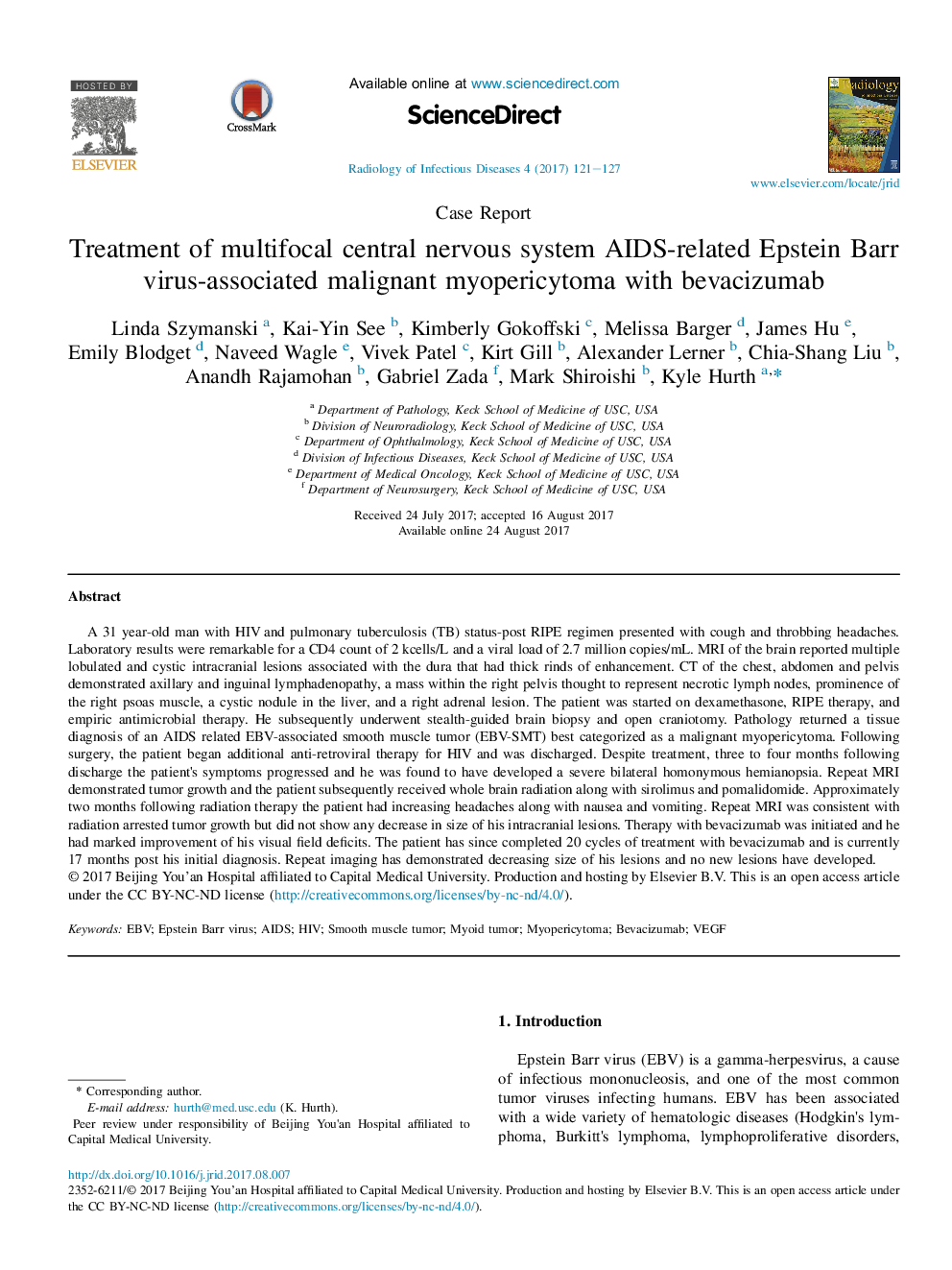
A 31 year-old man with HIV and pulmonary tuberculosis (TB) status-post RIPE regimen presented with cough and throbbing headaches. Laboratory results were remarkable for a CD4 count of 2Â kcells/L and a viral load of 2.7 million copies/mL. MRI of the brain reported multiple lobulated and cystic intracranial lesions associated with the dura that had thick rinds of enhancement. CT of the chest, abdomen and pelvis demonstrated axillary and inguinal lymphadenopathy, a mass within the right pelvis thought to represent necrotic lymph nodes, prominence of the right psoas muscle, a cystic nodule in the liver, and a right adrenal lesion. The patient was started on dexamethasone, RIPE therapy, and empiric antimicrobial therapy. He subsequently underwent stealth-guided brain biopsy and open craniotomy. Pathology returned a tissue diagnosis of an AIDS related EBV-associated smooth muscle tumor (EBV-SMT) best categorized as a malignant myopericytoma. Following surgery, the patient began additional anti-retroviral therapy for HIV and was discharged. Despite treatment, three to four months following discharge the patient's symptoms progressed and he was found to have developed a severe bilateral homonymous hemianopsia. Repeat MRI demonstrated tumor growth and the patient subsequently received whole brain radiation along with sirolimus and pomalidomide. Approximately two months following radiation therapy the patient had increasing headaches along with nausea and vomiting. Repeat MRI was consistent with radiation arrested tumor growth but did not show any decrease in size of his intracranial lesions. Therapy with bevacizumab was initiated and he had marked improvement of his visual field deficits. The patient has since completed 20 cycles of treatment with bevacizumab and is currently 17 months post his initial diagnosis. Repeat imaging has demonstrated decreasing size of his lesions and no new lesions have developed.
ناشر
Database: Elsevier - ScienceDirect (ساینس دایرکت)
Journal: Radiology of Infectious Diseases - Volume 4, Issue 3, September 2017, Pages 121-127
Journal: Radiology of Infectious Diseases - Volume 4, Issue 3, September 2017, Pages 121-127
نویسندگان
Linda Szymanski, Kai-Yin See, Kimberly Gokoffski, Melissa Barger, James Hu, Emily Blodget, Naveed Wagle, Vivek Patel, Kirt Gill, Alexander Lerner, Chia-Shang Liu, Anandh Rajamohan, Gabriel Zada, Mark Shiroishi, Kyle Hurth,