| کد مقاله | کد نشریه | سال انتشار | مقاله انگلیسی | نسخه تمام متن |
|---|---|---|---|---|
| 8735457 | 1591048 | 2016 | 5 صفحه PDF | دانلود رایگان |
عنوان انگلیسی مقاله ISI
Hepatic perivascular epithelioid cell tumor in three patients
ترجمه فارسی عنوان
تومور سلول های اپیتلیویید پریواسکولار در سه بیمار
دانلود مقاله + سفارش ترجمه
دانلود مقاله ISI انگلیسی
رایگان برای ایرانیان
موضوعات مرتبط
علوم پزشکی و سلامت
پزشکی و دندانپزشکی
کبدشناسی
چکیده انگلیسی
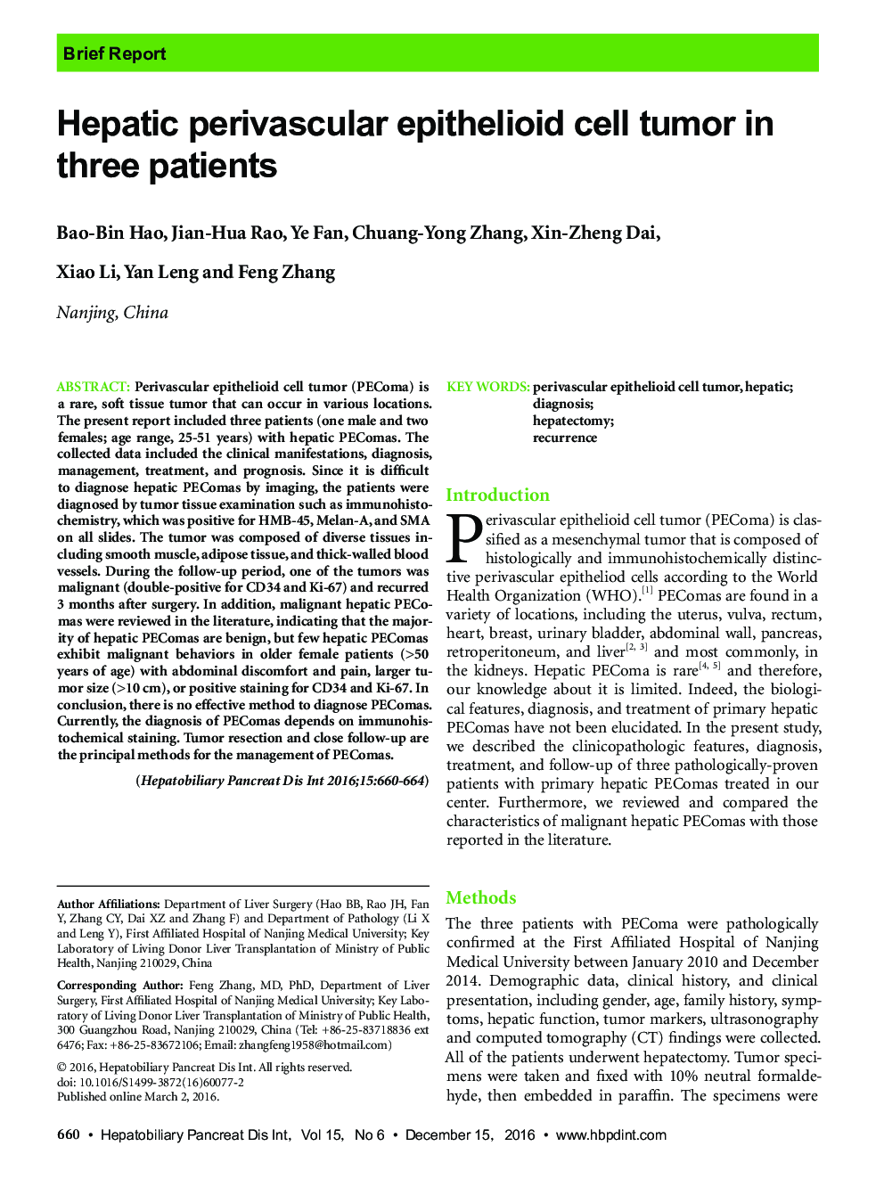
Perivascular epithelioid cell tumor (PEComa) is a rare, soft tissue tumor that can occur in various locations. The present report included three patients (one male and two females; age range, 25-51 years) with hepatic PEComas. The collected data included the clinical manifestations, diagnosis, management, treatment, and prognosis. Since it is difficult to diagnose hepatic PEComas by imaging, the patients were diagnosed by tumor tissue examination such as immunohistochemistry, which was positive for HMB-45, Melan-A, and SMA on all slides. The tumor was composed of diverse tissues including smooth muscle, adipose tissue, and thick-walled blood vessels. During the follow-up period, one of the tumors was malignant (double-positive for CD34 and Ki-67) and recurred 3 months after surgery. In addition, malignant hepatic PEComas were reviewed in the literature, indicating that the majority of hepatic PEComas are benign, but few hepatic PEComas exhibit malignant behaviors in older female patients (>50 years of age) with abdominal discomfort and pain, larger tumor size (>10 cm), or positive staining for CD34 and Ki-67. In conclusion, there is no effective method to diagnose PEComas. Currently, the diagnosis of PEComas depends on immunohistochemical staining. Tumor resection and close follow-up are the principal methods for the management of PEComas.
ناشر
Database: Elsevier - ScienceDirect (ساینس دایرکت)
Journal: Hepatobiliary & Pancreatic Diseases International - Volume 15, Issue 6, 15 December 2016, Pages 660-664
Journal: Hepatobiliary & Pancreatic Diseases International - Volume 15, Issue 6, 15 December 2016, Pages 660-664
نویسندگان
Bao-Bin Hao, Jian-Hua Rao, Ye Fan, Chuang-Yong Zhang, Xin-Zheng Dai, Xiao Li, Yan Leng, Feng Zhang,
