| کد مقاله | کد نشریه | سال انتشار | مقاله انگلیسی | نسخه تمام متن |
|---|---|---|---|---|
| 8996621 | 1115138 | 2005 | 8 صفحه PDF | دانلود رایگان |
عنوان انگلیسی مقاله ISI
T waves are independently generated in both right and left ventricles: The clinically recorded T is a summation of two separate repolarizations, and is thus always biventricular
دانلود مقاله + سفارش ترجمه
دانلود مقاله ISI انگلیسی
رایگان برای ایرانیان
موضوعات مرتبط
علوم زیستی و بیوفناوری
بیوشیمی، ژنتیک و زیست شناسی مولکولی
زیست شناسی تکاملی
پیش نمایش صفحه اول مقاله

چکیده انگلیسی
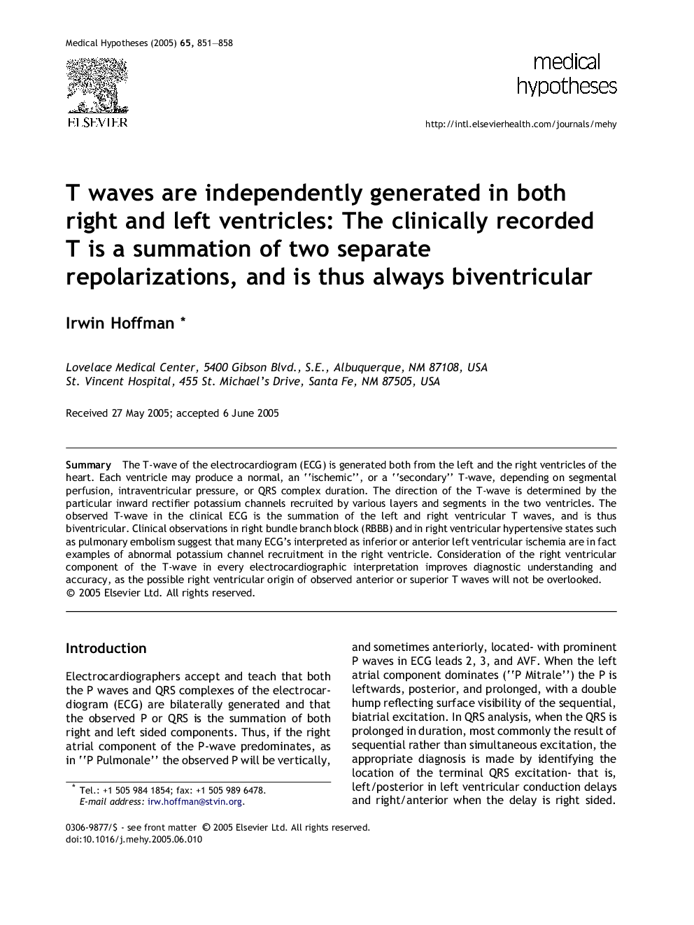
The T-wave of the electrocardiogram (ECG) is generated both from the left and the right ventricles of the heart. Each ventricle may produce a normal, an “ischemic”, or a “secondary” T-wave, depending on segmental perfusion, intraventricular pressure, or QRS complex duration. The direction of the T-wave is determined by the particular inward rectifier potassium channels recruited by various layers and segments in the two ventricles. The observed T-wave in the clinical ECG is the summation of the left and right ventricular T waves, and is thus biventricular. Clinical observations in right bundle branch block (RBBB) and in right ventricular hypertensive states such as pulmonary embolism suggest that many ECG's interpreted as inferior or anterior left ventricular ischemia are in fact examples of abnormal potassium channel recruitment in the right ventricle. Consideration of the right ventricular component of the T-wave in every electrocardiographic interpretation improves diagnostic understanding and accuracy, as the possible right ventricular origin of observed anterior or superior T waves will not be overlooked.
ناشر
Database: Elsevier - ScienceDirect (ساینس دایرکت)
Journal: Medical Hypotheses - Volume 65, Issue 5, 2005, Pages 851-858
Journal: Medical Hypotheses - Volume 65, Issue 5, 2005, Pages 851-858
نویسندگان
Irwin Hoffman,