| کد مقاله | کد نشریه | سال انتشار | مقاله انگلیسی | نسخه تمام متن |
|---|---|---|---|---|
| 9021138 | 1561376 | 2005 | 5 صفحه PDF | دانلود رایگان |
عنوان انگلیسی مقاله ISI
3D-rotational X-ray imaging (3D-RX) for the depiction of the trabecular bone structure
دانلود مقاله + سفارش ترجمه
دانلود مقاله ISI انگلیسی
رایگان برای ایرانیان
کلمات کلیدی
موضوعات مرتبط
علوم زیستی و بیوفناوری
بیوشیمی، ژنتیک و زیست شناسی مولکولی
زیست شناسی مولکولی
پیش نمایش صفحه اول مقاله

چکیده انگلیسی
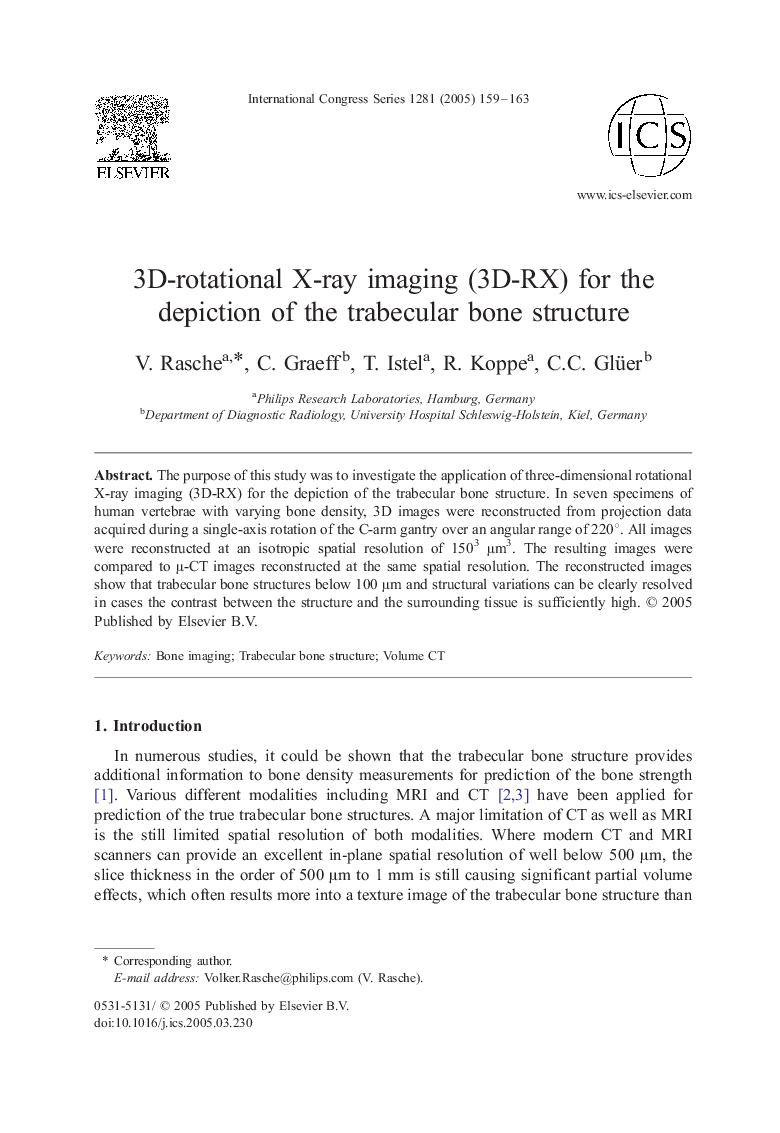
The purpose of this study was to investigate the application of three-dimensional rotational X-ray imaging (3D-RX) for the depiction of the trabecular bone structure. In seven specimens of human vertebrae with varying bone density, 3D images were reconstructed from projection data acquired during a single-axis rotation of the C-arm gantry over an angular range of 220°. All images were reconstructed at an isotropic spatial resolution of 1503 μm3. The resulting images were compared to μ-CT images reconstructed at the same spatial resolution. The reconstructed images show that trabecular bone structures below 100 μm and structural variations can be clearly resolved in cases the contrast between the structure and the surrounding tissue is sufficiently high.
ناشر
Database: Elsevier - ScienceDirect (ساینس دایرکت)
Journal: International Congress Series - Volume 1281, May 2005, Pages 159-163
Journal: International Congress Series - Volume 1281, May 2005, Pages 159-163
نویسندگان
V. Rasche, C. Graeff, T. Istel, R. Koppe, C.C. Glüer,