| کد مقاله | کد نشریه | سال انتشار | مقاله انگلیسی | نسخه تمام متن |
|---|---|---|---|---|
| 9088326 | 1148077 | 2005 | 33 صفحه PDF | دانلود رایگان |
عنوان انگلیسی مقاله ISI
Magnetic Resonance Imaging in the Characterization of Pelvic Masses
دانلود مقاله + سفارش ترجمه
دانلود مقاله ISI انگلیسی
رایگان برای ایرانیان
موضوعات مرتبط
علوم پزشکی و سلامت
پزشکی و دندانپزشکی
رادیولوژی و تصویربرداری
پیش نمایش صفحه اول مقاله

چکیده انگلیسی
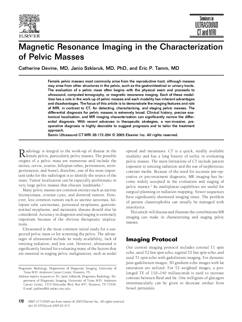
Female pelvic masses most commonly arise from the reproductive tract, although masses may arise from other structures in the pelvis, such as the gastrointestinal or urinary tracts. The evaluation of a pelvic mass often begins with the physical exam and proceeds to ultrasound, computed tomography, or magnetic resonance imaging. Each of these modalities has a role in the work-up of pelvic masses and each modality has inherent advantages and disadvantages. The focus of this article is to demonstrate the imaging features and role of MRI, in contrast to CT, for detecting, characterizing, and staging pelvic masses. The differential diagnosis for pelvic masses is extremely broad. Clinical history, precise anatomical localization, and MR imaging characterization can significantly narrow the differential diagnosis. With recent advances in therapeutic strategies, a non-invasive, pre-operative diagnosis is highly desirable to suggest prognosis and to tailor the treatment approach.
ناشر
Database: Elsevier - ScienceDirect (ساینس دایرکت)
Journal: Seminars in Ultrasound, CT and MRI - Volume 26, Issue 3, June 2005, Pages 172-204
Journal: Seminars in Ultrasound, CT and MRI - Volume 26, Issue 3, June 2005, Pages 172-204
نویسندگان
Catherine MD, Janio MD, PhD, Eric P. MD,