| کد مقاله | کد نشریه | سال انتشار | مقاله انگلیسی | نسخه تمام متن |
|---|---|---|---|---|
| 9245023 | 1209940 | 2005 | 10 صفحه PDF | دانلود رایگان |
عنوان انگلیسی مقاله ISI
Real-time imaging of human cortical activity evoked by painful esophageal stimulation
دانلود مقاله + سفارش ترجمه
دانلود مقاله ISI انگلیسی
رایگان برای ایرانیان
کلمات کلیدی
VASFGDROISAMCortical evoked potentialCEP - MOBILEFunctional gastrointestinal disorder - اختلال عملکرد دستگاه گوارشMEG - بهPositron emission tomography - توموگرافی گسیل پوزیترونSynthetic aperture magnetometry - مغناطیس سنج دیافراگم مصنوعیmagnetoencephalography - مغناطیس مغزvisual analogue scale - مقیاس آنالوگ بصریregion of interest - منطقه مورد نظرPET - پت
موضوعات مرتبط
علوم پزشکی و سلامت
پزشکی و دندانپزشکی
بیماریهای گوارشی
پیش نمایش صفحه اول مقاله

چکیده انگلیسی
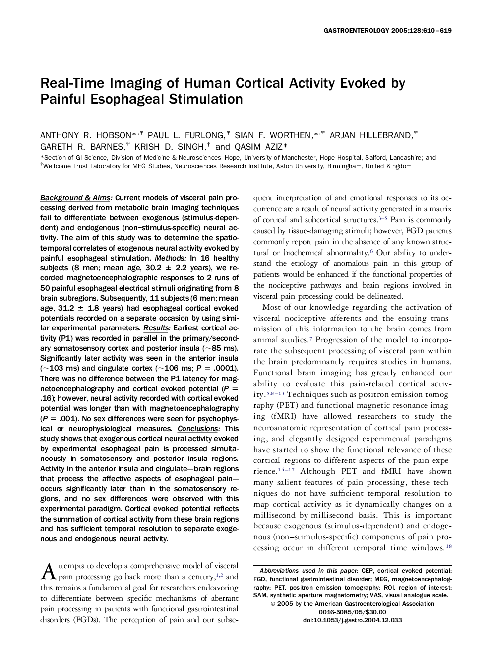
Background & aims: Current models of visceral pain processing derived from metabolic brain imaging techniques fail to differentiate between exogenous (stimulus-dependent) and endogenous (non-stimulus-specific) neural activity. The aim of this study was to determine the spatiotemporal correlates of exogenous neural activity evoked by painful esophageal stimulation. Methods: In 16 healthy subjects (8 men; mean age, 30.2 ± 2.2 years), we recorded magnetoencephalographic responses to 2 runs of 50 painful esophageal electrical stimuli originating from 8 brain subregions. Subsequently, 11 subjects (6 men; mean age, 31.2 ± 1.8 years) had esophageal cortical evoked potentials recorded on a separate occasion by using similar experimental parameters. Results: Earliest cortical activity (P1) was recorded in parallel in the primary/secondary somatosensory cortex and posterior insula (â¼85 ms). Significantly later activity was seen in the anterior insula (â¼103 ms) and cingulate cortex (â¼106 ms; P = .0001). There was no difference between the P1 latency for magnetoencephalography and cortical evoked potential (P = .16); however, neural activity recorded with cortical evoked potential was longer than with magnetoencephalography (P = .001). No sex differences were seen for psychophysical or neurophysiological measures. Conclusions: This study shows that exogenous cortical neural activity evoked by experimental esophageal pain is processed simultaneously in somatosensory and posterior insula regions. Activity in the anterior insula and cingulate-brain regions that process the affective aspects of esophageal pain-occurs significantly later than in the somatosensory regions, and no sex differences were observed with this experimental paradigm. Cortical evoked potential reflects the summation of cortical activity from these brain regions and has sufficient temporal resolution to separate exogenous and endogenous neural activity.
ناشر
Database: Elsevier - ScienceDirect (ساینس دایرکت)
Journal: Gastroenterology - Volume 128, Issue 3, March 2005, Pages 610-619
Journal: Gastroenterology - Volume 128, Issue 3, March 2005, Pages 610-619
نویسندگان
Anthony R. Hobson, Paul L. Furlong, Sian F. Worthen, Arjan Hillebrand, Gareth R. Barnes, Krish D. Singh, Qasim Aziz,