| کد مقاله | کد نشریه | سال انتشار | مقاله انگلیسی | نسخه تمام متن |
|---|---|---|---|---|
| 9345737 | 1262369 | 2005 | 4 صفحه PDF | دانلود رایگان |
عنوان انگلیسی مقاله ISI
Difficultés diagnostiques face à une tumeur papillaire pigmentée. à propos d'un cas
دانلود مقاله + سفارش ترجمه
دانلود مقاله ISI انگلیسی
رایگان برای ایرانیان
موضوعات مرتبط
علوم پزشکی و سلامت
پزشکی و دندانپزشکی
چشم پزشکی
پیش نمایش صفحه اول مقاله

چکیده انگلیسی
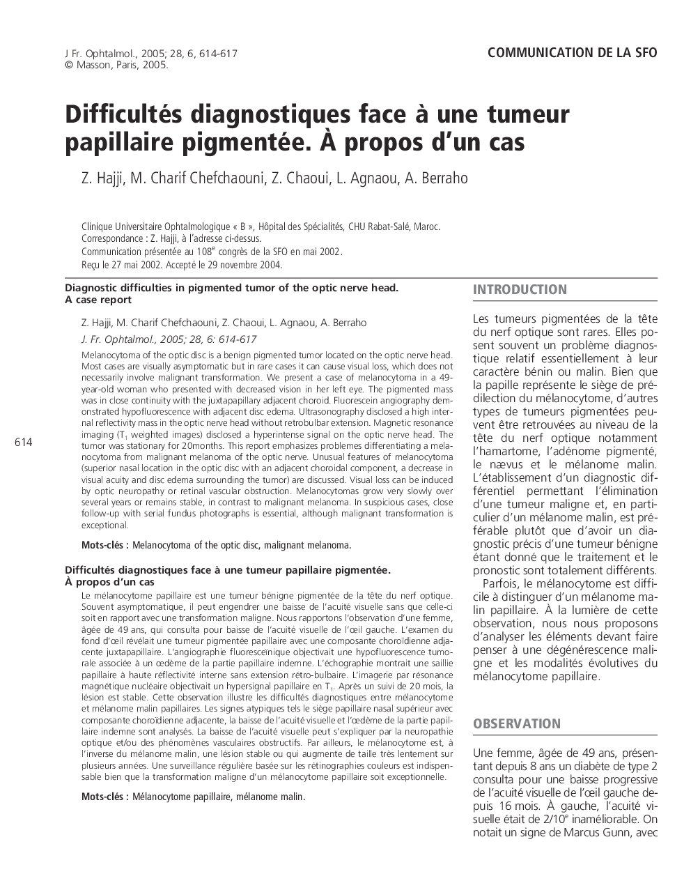
Melanocytoma of the optic disc is a benign pigmented tumor located on the optic nerve head. Most cases are visually asymptomatic but in rare cases it can cause visual loss, which does not necessarily involve malignant transformation. We present a case of melanocytoma in a 49-year-old woman who presented with decreased vision in her left eye. The pigmented mass was in close continuity with the juxtapapillary adjacent choroid. Fluorescein angiography demonstrated hypofluorescence with adjacent disc edema. Ultrasonography disclosed a high internal reflectivity mass in the optic nerve head without retrobulbar extension. Magnetic resonance imaging (T1 weighted images) disclosed a hyperintense signal on the optic nerve head. The tumor was stationary for 20months. This report emphasizes problemes differentiating a melanocytoma from malignant melanoma of the optic nerve. Unusual features of melanocytoma (superior nasal location in the optic disc with an adjacent choroidal component, a decrease in visual acuity and disc edema surrounding the tumor) are discussed. Visual loss can be induced by optic neuropathy or retinal vascular obstruction. Melanocytomas grow very slowly over several years or remains stable, in contrast to malignant melanoma. In suspicious cases, close follow-up with serial fundus photographs is essential, although malignant transformation is exceptional.
ناشر
Database: Elsevier - ScienceDirect (ساینس دایرکت)
Journal: Journal Français d'Ophtalmologie - Volume 28, Issue 6, June 2005, Pages 614-617
Journal: Journal Français d'Ophtalmologie - Volume 28, Issue 6, June 2005, Pages 614-617
نویسندگان
Z. Hajji, M. Charif Chefchaouni, Z. Chaoui, L. Agnaou, A. Berraho,