| کد مقاله | کد نشریه | سال انتشار | مقاله انگلیسی | نسخه تمام متن |
|---|---|---|---|---|
| 9345824 | 1262376 | 2005 | 11 صفحه PDF | دانلود رایگان |
عنوان انگلیسی مقاله ISI
Intérêt de la microscopie confocale in vitro dans l'étude des membranes épirétiniennes et des membranes limitantes internes après chirurgie maculaire
دانلود مقاله + سفارش ترجمه
دانلود مقاله ISI انگلیسی
رایگان برای ایرانیان
کلمات کلیدی
موضوعات مرتبط
علوم پزشکی و سلامت
پزشکی و دندانپزشکی
چشم پزشکی
پیش نمایش صفحه اول مقاله

چکیده انگلیسی
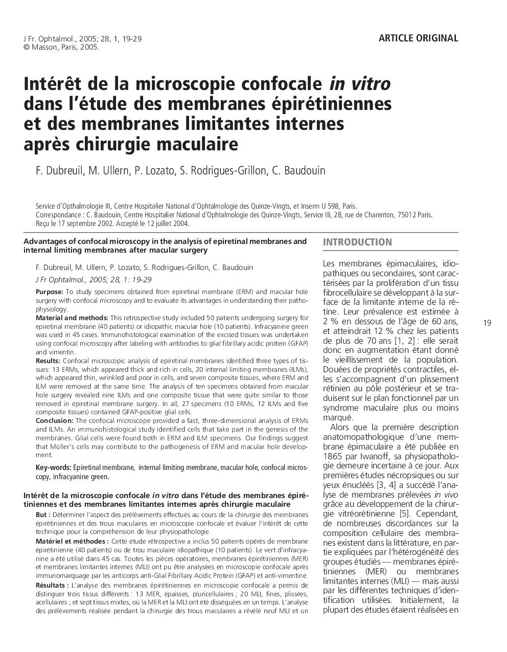
The confocal microscope provided a fast, three-dimensional analysis of ERMs and ILMs. An immunohistological study identified cells that take part in the genesis of the membranes. Glial cells were found both in ERM and ILM specimens. Our findings suggest that Müller's cells may contribute to the pathogenesis of ERM and macular hole development.
ناشر
Database: Elsevier - ScienceDirect (ساینس دایرکت)
Journal: Journal Français d'Ophtalmologie - Volume 28, Issue 1, January 2005, Pages 19-29
Journal: Journal Français d'Ophtalmologie - Volume 28, Issue 1, January 2005, Pages 19-29
نویسندگان
F. Dubreuil, M. Ullern, P. Lozato, S. Rodrigues-Grillon, C. Baudouin,