| کد مقاله | کد نشریه | سال انتشار | مقاله انگلیسی | نسخه تمام متن |
|---|---|---|---|---|
| 9345985 | 1262386 | 2005 | 8 صفحه PDF | دانلود رایگان |
عنوان انگلیسی مقاله ISI
Microscopie confocale et pathologies de la surface oculaire : corrélations anatomo-cliniques
دانلود مقاله + سفارش ترجمه
دانلود مقاله ISI انگلیسی
رایگان برای ایرانیان
کلمات کلیدی
موضوعات مرتبط
علوم پزشکی و سلامت
پزشکی و دندانپزشکی
چشم پزشکی
پیش نمایش صفحه اول مقاله

چکیده انگلیسی
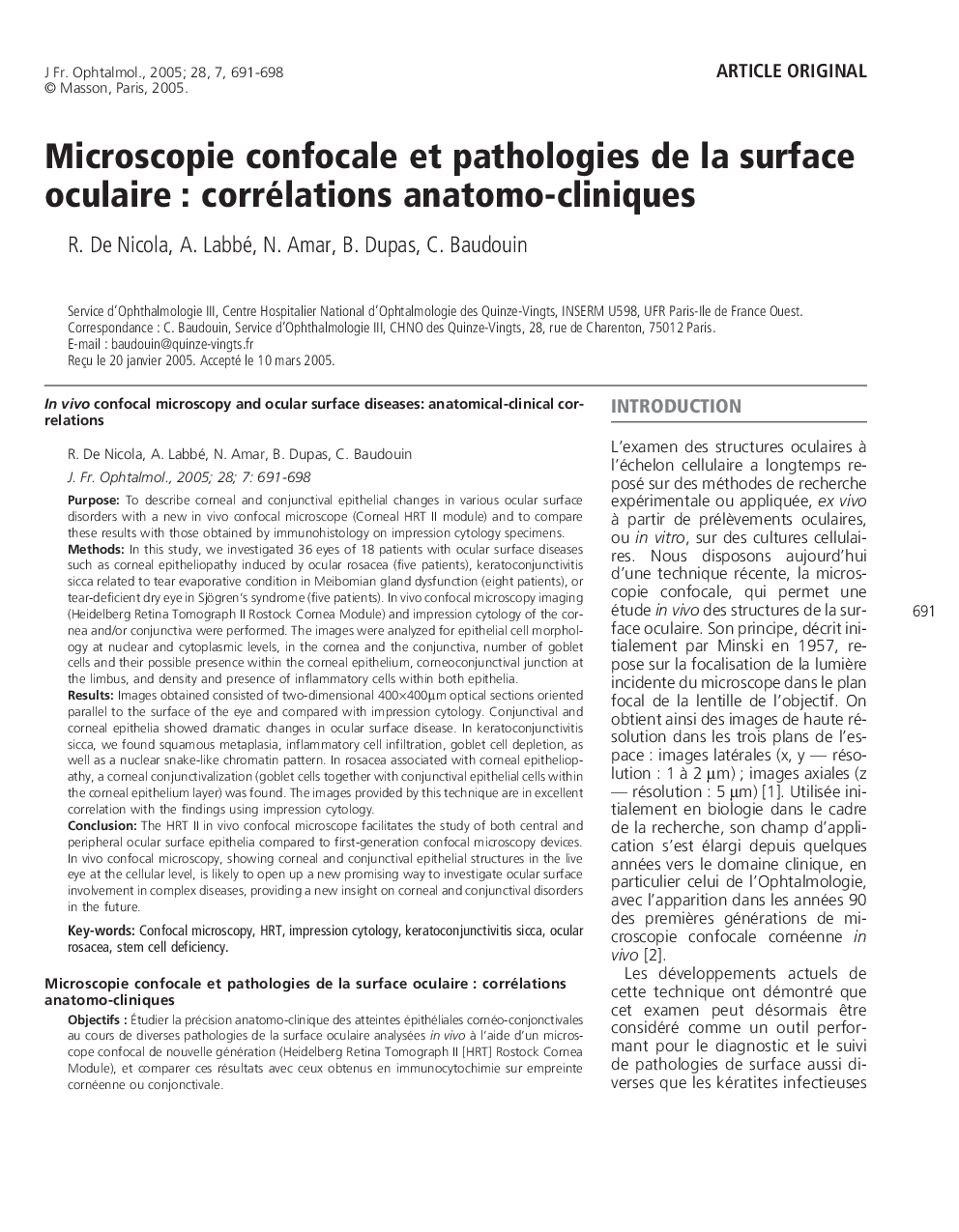
The HRT II in vivo confocal microscope facilitates the study of both central and peripheral ocular surface epithelia compared to first-generation confocal microscopy devices. In vivo confocal microscopy, showing corneal and conjunctival epithelial structures in the live eye at the cellular level, is likely to open up a new promising way to investigate ocular surface involvement in complex diseases, providing a new insight on corneal and conjunctival disorders in the future.
ناشر
Database: Elsevier - ScienceDirect (ساینس دایرکت)
Journal: Journal Français d'Ophtalmologie - Volume 28, Issue 7, September 2005, Pages 691-698
Journal: Journal Français d'Ophtalmologie - Volume 28, Issue 7, September 2005, Pages 691-698
نویسندگان
R. De Nicola, A. Labbé, N. Amar, B. Dupas, C. Baudouin,