| کد مقاله | کد نشریه | سال انتشار | مقاله انگلیسی | نسخه تمام متن |
|---|---|---|---|---|
| 9365767 | 1271524 | 2005 | 4 صفحه PDF | دانلود رایگان |
عنوان انگلیسی مقاله ISI
Subepidermal cleft formation as a diagnostic marker for cutaneous malignant melanoma
دانلود مقاله + سفارش ترجمه
دانلود مقاله ISI انگلیسی
رایگان برای ایرانیان
کلمات کلیدی
موضوعات مرتبط
علوم پزشکی و سلامت
پزشکی و دندانپزشکی
آسیبشناسی و فناوری پزشکی
پیش نمایش صفحه اول مقاله

چکیده انگلیسی
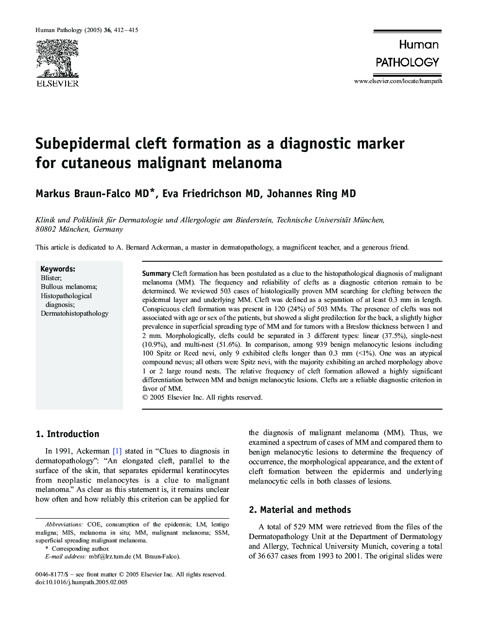
Cleft formation has been postulated as a clue to the histopathological diagnosis of malignant melanoma (MM). The frequency and reliability of clefts as a diagnostic criterion remain to be determined. We reviewed 503 cases of histologically proven MM searching for clefting between the epidermal layer and underlying MM. Cleft was defined as a separation of at least 0.3 mm in length. Conspicuous cleft formation was present in 120 (24%) of 503 MMs. The presence of clefts was not associated with age or sex of the patients, but showed a slight predilection for the back, a slightly higher prevalence in superficial spreading type of MM and for tumors with a Breslow thickness between 1 and 2 mm. Morphologically, clefts could be separated in 3 different types: linear (37.5%), single-nest (10.9%), and multi-nest (51.6%). In comparison, among 939 benign melanocytic lesions including 100 Spitz or Reed nevi, only 9 exhibited clefts longer than 0.3 mm (<1%). One was an atypical compound nevus; all others were Spitz nevi, with the majority exhibiting an arched morphology above 1 or 2 large round nests. The relative frequency of cleft formation allowed a highly significant differentiation between MM and benign melanocytic lesions. Clefts are a reliable diagnostic criterion in favor of MM.
ناشر
Database: Elsevier - ScienceDirect (ساینس دایرکت)
Journal: Human Pathology - Volume 36, Issue 4, April 2005, Pages 412-415
Journal: Human Pathology - Volume 36, Issue 4, April 2005, Pages 412-415
نویسندگان
Markus MD, Eva MD, Johannes MD,