| کد مقاله | کد نشریه | سال انتشار | مقاله انگلیسی | نسخه تمام متن |
|---|---|---|---|---|
| 9425358 | 1295863 | 2005 | 23 صفحه PDF | دانلود رایگان |
عنوان انگلیسی مقاله ISI
Prolonged translation arrest in reperfused hippocampal cornu Ammonis 1 is mediated by stress granules
دانلود مقاله + سفارش ترجمه
دانلود مقاله ISI انگلیسی
رایگان برای ایرانیان
کلمات کلیدی
موضوعات مرتبط
علوم زیستی و بیوفناوری
علم عصب شناسی
علوم اعصاب (عمومی)
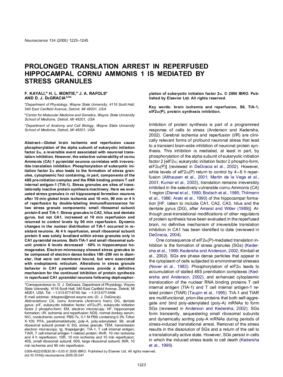
پیش نمایش صفحه اول مقاله

چکیده انگلیسی
Global brain ischemia and reperfusion cause phosphorylation of the alpha subunit of eukaryotic initiation factor 2α, a reversible event associated with neuronal translation inhibition. However, the selective vulnerability of cornu Ammonis (CA) 1 pyramidal neurons correlates with irreversible translation inhibition. Phosphorylation of eukaryotic initiation factor 2α also leads to the formation of stress granules, cytoplasmic foci containing, in part, components of the 48S pre-initiation complex and the RNA binding protein T cell internal antigen-1 (TIA-1). Stress granules are sites of translationally inactive protein synthesis machinery. Here we evaluated stress granules in rat hippocampal formation neurons after 10 min global brain ischemia and 10 min, 90 min or 4 h of reperfusion by double-labeling immunofluorescence for two stress granule components: small ribosomal subunit protein 6 and TIA-1. Stress granules in CA3, hilus and dentate gyrus, but not CA1, increased at 10 min reperfusion and returned to control levels by 90 min reperfusion. Dynamic changes in the nuclear distribution of TIA-1 occurred in resistant neurons. At 4 h reperfusion, small ribosomal subunit protein 6 was solely localized within stress granules only in CA1 pyramidal neurons. Both TIA-1 and small ribosomal subunit protein 6 levels decreased â¼50% in hippocampus homogenates. Electron microscopy showed stress granules to be composed of electron dense bodies 100-200nm in diameter, that were not membrane bound, but were associated with endoplasmic reticulum. Alterations in stress granule behavior in CA1 pyramidal neurons provide a definitive mechanism for the continued inhibition of protein synthesis in reperfused CA1 pyramidal neurons following dephosphorylation of eukaryotic initiation factor 2α.
ناشر
Database: Elsevier - ScienceDirect (ساینس دایرکت)
Journal: Neuroscience - Volume 134, Issue 4, 2005, Pages 1223-1245
Journal: Neuroscience - Volume 134, Issue 4, 2005, Pages 1223-1245
نویسندگان
F. Kayali, H.L. Montie, J.A. Rafols, D.J. DeGracia,