| کد مقاله | کد نشریه | سال انتشار | مقاله انگلیسی | نسخه تمام متن |
|---|---|---|---|---|
| 9872100 | 1533184 | 2005 | 6 صفحه PDF | دانلود رایگان |
عنوان انگلیسی مقاله ISI
Use of CD-ROM-based tool for analyzing contouring variations in involved-field radiotherapy for Stage III NSCLC
دانلود مقاله + سفارش ترجمه
دانلود مقاله ISI انگلیسی
رایگان برای ایرانیان
کلمات کلیدی
موضوعات مرتبط
مهندسی و علوم پایه
فیزیک و نجوم
تشعشع
پیش نمایش صفحه اول مقاله

چکیده انگلیسی
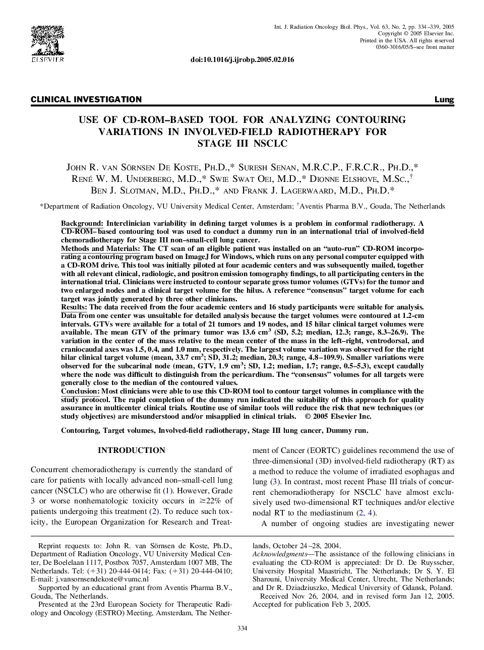
Background: Interclinician variability in defining target volumes is a problem in conformal radiotherapy. A CD-ROM-based contouring tool was used to conduct a dummy run in an international trial of involved-field chemoradiotherapy for Stage III non-small-cell lung cancer. Methods and Materials: The CT scan of an eligible patient was installed on an “auto-run” CD-ROM incorporating a contouring program based on ImageJ for Windows, which runs on any personal computer equipped with a CD-ROM drive. This tool was initially piloted at four academic centers and was subsequently mailed, together with all relevant clinical, radiologic, and positron emission tomography findings, to all participating centers in the international trial. Clinicians were instructed to contour separate gross tumor volumes (GTVs) for the tumor and two enlarged nodes and a clinical target volume for the hilus. A reference “consensus” target volume for each target was jointly generated by three other clinicians. Results: The data received from the four academic centers and 16 study participants were suitable for analysis. Data from one center was unsuitable for detailed analysis because the target volumes were contoured at 1.2-cm intervals. GTVs were available for a total of 21 tumors and 19 nodes, and 15 hilar clinical target volumes were available. The mean GTV of the primary tumor was 13.6 cm3 (SD, 5.2; median, 12.3; range, 8.3-26.9). The variation in the center of the mass relative to the mean center of the mass in the left-right, ventrodorsal, and craniocaudal axes was 1.5, 0.4, and 1.0 mm, respectively. The largest volume variation was observed for the right hilar clinical target volume (mean, 33.7 cm3; SD, 31.2; median, 20.3; range, 4.8-109.9). Smaller variations were observed for the subcarinal node (mean, GTV, 1.9 cm3; SD, 1.2; median, 1.7; range, 0.5-5.3), except caudally where the node was difficult to distinguish from the pericardium. The “consensus” volumes for all targets were generally close to the median of the contoured values. Conclusion: Most clinicians were able to use this CD-ROM tool to contour target volumes in compliance with the study protocol. The rapid completion of the dummy run indicated the suitability of this approach for quality assurance in multicenter clinical trials. Routine use of similar tools will reduce the risk that new techniques (or study objectives) are misunderstood and/or misapplied in clinical trials.
ناشر
Database: Elsevier - ScienceDirect (ساینس دایرکت)
Journal: International Journal of Radiation Oncology*Biology*Physics - Volume 63, Issue 2, 1 October 2005, Pages 334-339
Journal: International Journal of Radiation Oncology*Biology*Physics - Volume 63, Issue 2, 1 October 2005, Pages 334-339
نویسندگان
John R. Ph.D., Suresh (F.R.C.R.), René W.M. M.D., Swie Swat M.D., Dionne M.Sc., Ben J. M.D., Ph.D., Frank J. M.D., Ph.D.,