| کد مقاله | کد نشریه | سال انتشار | مقاله انگلیسی | نسخه تمام متن |
|---|---|---|---|---|
| 10078737 | 1603616 | 2005 | 10 صفحه PDF | دانلود رایگان |
عنوان انگلیسی مقاله ISI
Use of Preoperative Magnetic Resonance Imaging to Predict Rotator Cuff Tear Pattern and Method of Repair
دانلود مقاله + سفارش ترجمه
دانلود مقاله ISI انگلیسی
رایگان برای ایرانیان
کلمات کلیدی
موضوعات مرتبط
علوم پزشکی و سلامت
پزشکی و دندانپزشکی
ارتوپدی، پزشکی ورزشی و توانبخشی
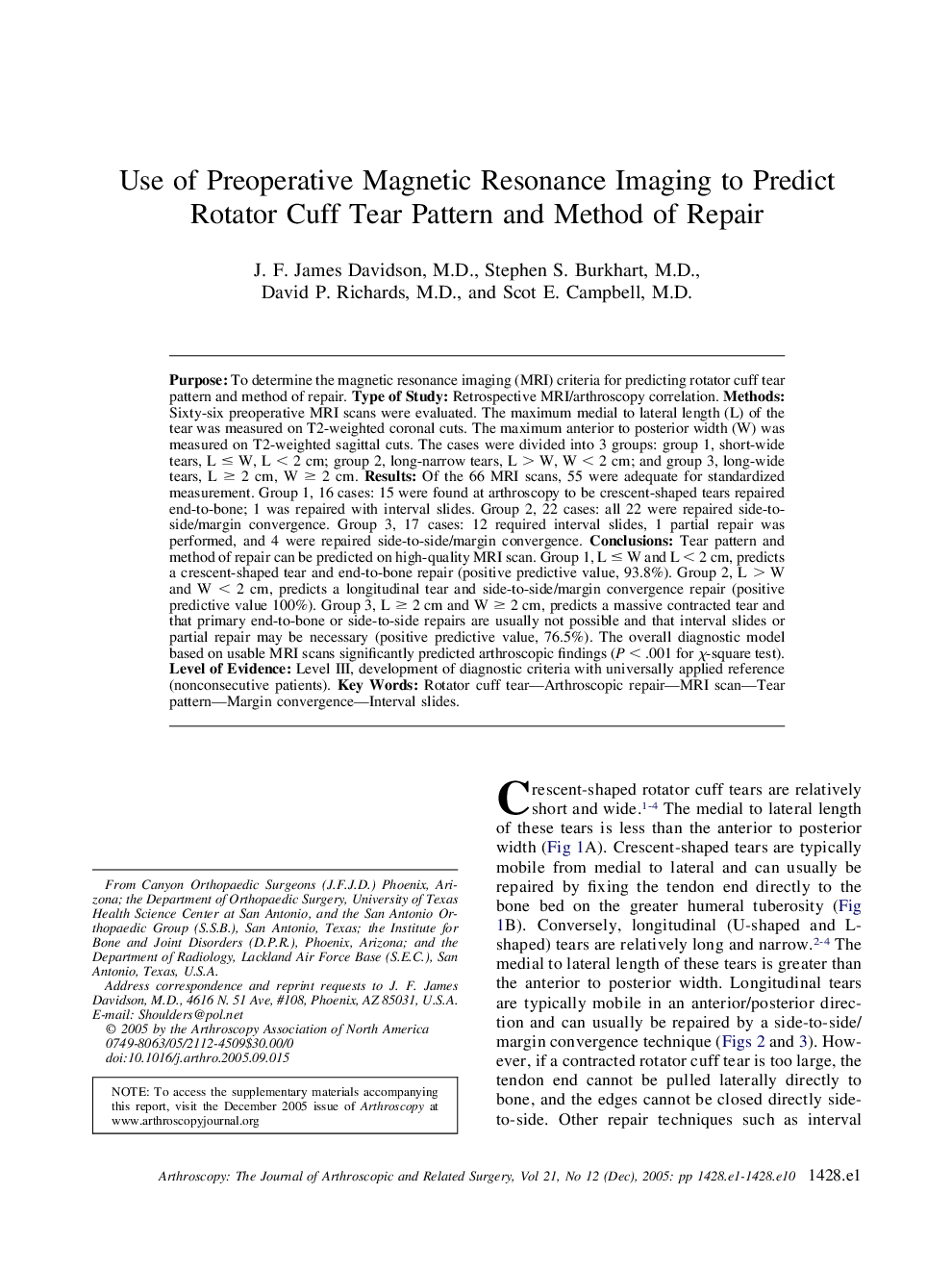
پیش نمایش صفحه اول مقاله

چکیده انگلیسی
Purpose: To determine the magnetic resonance imaging (MRI) criteria for predicting rotator cuff tear pattern and method of repair. Type of Study: Retrospective MRI/arthroscopy correlation. Methods: Sixty-six preoperative MRI scans were evaluated. The maximum medial to lateral length (L) of the tear was measured on T2-weighted coronal cuts. The maximum anterior to posterior width (W) was measured on T2-weighted sagittal cuts. The cases were divided into 3 groups: group 1, short-wide tears, L ⤠W, L < 2 cm; group 2, long-narrow tears, L > W, W < 2 cm; and group 3, long-wide tears, L ⥠2 cm, W ⥠2 cm. Results: Of the 66 MRI scans, 55 were adequate for standardized measurement. Group 1, 16 cases: 15 were found at arthroscopy to be crescent-shaped tears repaired end-to-bone; 1 was repaired with interval slides. Group 2, 22 cases: all 22 were repaired side-to-side/margin convergence. Group 3, 17 cases: 12 required interval slides, 1 partial repair was performed, and 4 were repaired side-to-side/margin convergence. Conclusions: Tear pattern and method of repair can be predicted on high-quality MRI scan. Group 1, L ⤠W and L < 2 cm, predicts a crescent-shaped tear and end-to-bone repair (positive predictive value, 93.8%). Group 2, L > W and W < 2 cm, predicts a longitudinal tear and side-to-side/margin convergence repair (positive predictive value 100%). Group 3, L ⥠2 cm and W ⥠2 cm, predicts a massive contracted tear and that primary end-to-bone or side-to-side repairs are usually not possible and that interval slides or partial repair may be necessary (positive predictive value, 76.5%). The overall diagnostic model based on usable MRI scans significantly predicted arthroscopic findings (P < .001 for Ï-square test). Level of Evidence: Level III, development of diagnostic criteria with universally applied reference (nonconsecutive patients).
ناشر
Database: Elsevier - ScienceDirect (ساینس دایرکت)
Journal: Arthroscopy: The Journal of Arthroscopic & Related Surgery - Volume 21, Issue 12, December 2005, Pages 1428.e1-1428.e10
Journal: Arthroscopy: The Journal of Arthroscopic & Related Surgery - Volume 21, Issue 12, December 2005, Pages 1428.e1-1428.e10
نویسندگان
J.F. M.D., Stephen S. M.D., David P. M.D., Scot E. M.D.,