| کد مقاله | کد نشریه | سال انتشار | مقاله انگلیسی | نسخه تمام متن |
|---|---|---|---|---|
| 10211546 | 1668026 | 2018 | 4 صفحه PDF | دانلود رایگان |
عنوان انگلیسی مقاله ISI
Comparison of Premortem Magnetic Resonance Imaging and Postmortem Autopsy Findings of a Cortical Microinfarct
دانلود مقاله + سفارش ترجمه
دانلود مقاله ISI انگلیسی
رایگان برای ایرانیان
کلمات کلیدی
موضوعات مرتبط
علوم پزشکی و سلامت
پزشکی و دندانپزشکی
مغز و اعصاب بالینی
پیش نمایش صفحه اول مقاله

چکیده انگلیسی
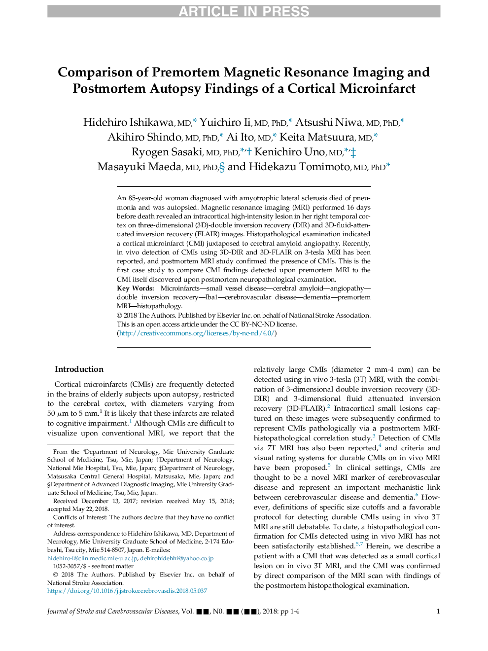
An 85-year-old woman diagnosed with amyotrophic lateral sclerosis died of pneumonia and was autopsied. Magnetic resonance imaging (MRI) performed 16 days before death revealed an intracortical high-intensity lesion in her right temporal cortex on three-dimensional (3D)-double inversion recovery (DIR) and 3D-fluid-attenuated inversion recovery (FLAIR) images. Histopathological examination indicated a cortical microinfarct (CMI) juxtaposed to cerebral amyloid angiopathy. Recently, in vivo detection of CMIs using 3D-DIR and 3D-FLAIR on 3-tesla MRI has been reported, and postmortem MRI study confirmed the presence of CMIs. This is the first case study to compare CMI findings detected upon premortem MRI to the CMI itself discovered upon postmortem neuropathological examination.
ناشر
Database: Elsevier - ScienceDirect (ساینس دایرکت)
Journal: Journal of Stroke and Cerebrovascular Diseases - Volume 27, Issue 10, October 2018, Pages 2623-2626
Journal: Journal of Stroke and Cerebrovascular Diseases - Volume 27, Issue 10, October 2018, Pages 2623-2626
نویسندگان
Hidehiro MD, Yuichiro MD, PhD, Atsushi MD, PhD, Akihiro MD, PhD, Ai MD, Keita MD, Ryogen MD, PhD, Kenichiro MD, Masayuki MD, PhD, Hidekazu MD, PhD,