| کد مقاله | کد نشریه | سال انتشار | مقاله انگلیسی | نسخه تمام متن |
|---|---|---|---|---|
| 10743418 | 1047888 | 2014 | 10 صفحه PDF | دانلود رایگان |
عنوان انگلیسی مقاله ISI
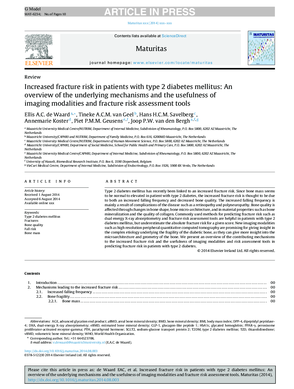
Increased fracture risk in patients with type 2 diabetes mellitus: An overview of the underlying mechanisms and the usefulness of imaging modalities and fracture risk assessment tools
ترجمه فارسی عنوان
افزایش ریسک شکستگی در بیماران مبتلا به دیابت نوع 2: یک مرور کلی از مکانیزم های زیربنایی و مفید بودن روش های تصویربرداری و ابزار ارزیابی ریسک شکست
دانلود مقاله + سفارش ترجمه
دانلود مقاله ISI انگلیسی
رایگان برای ایرانیان
کلمات کلیدی
PPAR-γSGLT2DPP-4BMDGLP-1pTHDXAaBMDHbA1cBone mineral density - تراکم معدنی استخوانAreal bone mineral density - تراکم معدنی استخوان منطقهdual-energy X-ray absorptiometry - جذب اندازه گیری اشعه ایکس دوگانه انرژیDipeptidyl peptidase-4 - دیپپتیدیل پپتیداز 4Age - سنbody mass index - شاخص توده بدنBMI - شاخص توده بدنیGlycated hemoglobin - هموگلوبین گلیکوزیلهparathyroid hormone - هورمون پاراتیروئیدperoxisome proliferator-activated receptor-gamma - پراکسیزوم پرولیفراتور فعال گیرنده گاماglucagon-like peptide 1 - پپتید مشابه گلوکاگون 1advanced glycation end product - پیشرفته محصول نهایی گلیساسیون
ترجمه چکیده
دیابت نوع 2 به تازگی با افزایش خطر شکستگی ارتباط دارد. از آنجاییکه توده استخوانی به نظر میرسد که در بیمار مبتلا به دیابت نوع 2 طبیعی است، خطر افزایش شکستگی ناشی از افزایش فراوانی کاهش و کاهش کیفیت استخوان است. فرکانس کاهش یافته به طور عمده نتیجه عوارض بیماری مانند رتینوپاتی و پلی یروپاتی است. کیفیت استخوان تحت تاثیر تغییرات در شکل استخوانی، معماری میکرو استخوان و خواص مواد مانند کانی سازی استخوان و کیفیت کلاژن است. روشهای رایج برای پیش بینی خطر شکستگی از قبیل انرژی جذب دوزهای انرژی دوگانه و ابزارهای ارزیابی خطر شکستگی در بیماران مبتلا به دیابت نوع 2 مفید هستند اما خطر شکست مطلق برای نمره داده شده را کم اهمیت می دانند. روشهای جدید تصویربرداری مانند توزیع باکتریایی با وضوح محیطی با وضوح بالا برای ارائه بینش در علت پیچیده اساسی شکننده بودن استخوان های دیابتی امیدوار کننده هستند، زیرا می توانند بینش های میکروارگانیسم و هندسه استخوان را بیشتر درک کنند. ما یک مرور کلی از مکانیسم های کمک کننده به افزایش ریسک شکست و سودمندی روش های تصویربرداری و ابزارهای ارزیابی خطر در پیش بینی خطر شکستگی در بیماران مبتلا به دیابت نوع 2 ارائه می دهیم.
موضوعات مرتبط
علوم زیستی و بیوفناوری
بیوشیمی، ژنتیک و زیست شناسی مولکولی
سالمندی
چکیده انگلیسی
Type 2 diabetes mellitus has recently been linked to an increased fracture risk. Since bone mass seems to be normal to elevated in patient with type 2 diabetes, the increased fracture risk is thought to be due to both an increased falling frequency and decreased bone quality. The increased falling frequency is mainly a result of complications of the disease such as a retinopathy and polyneuropathy. Bone quality is affected through changes in bone shape, bone micro-architecture, and in material properties such as bone mineralization and the quality of collagen. Commonly used methods for predicting fracture risk such as dual energy X-ray absorptiometry and fracture risk assessment tools are helpful in patients with type 2 diabetes mellitus, but underestimate the absolute fracture risk for a given score. New imaging modalities such as high resolution peripheral quantitative computed tomography are promising for giving insight in the complex etiology underlying the fragility of the diabetic bone, as they can give more insight into the microarchitecture and geometry of the bone. We present an overview of the contributing mechanisms to the increased fracture risk and the usefulness of imaging modalities and risk assessment tools in predicting fracture risk in patients with type 2 diabetes.
ناشر
Database: Elsevier - ScienceDirect (ساینس دایرکت)
Journal: Maturitas - Volume 79, Issue 3, November 2014, Pages 265-274
Journal: Maturitas - Volume 79, Issue 3, November 2014, Pages 265-274
نویسندگان
Ellis A.C. de Waard, Tineke A.C.M. van Geel, Hans H.C.M. Savelberg, Annemarie Koster, Piet P.M.M. Geusens, Joop P.W. van den Bergh,
